

 PDF
PDF ePub
ePub Citation
 Print
Print


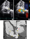
A 65-year-old female presented to the emergency room with fever and dyspnea for one week. The patient had a history of double aortic and mitral valve replacement 27 years prior to presentation and was started on warfarin since then. Four months before presentation the patient underwent spinal acupuncture which was complicated by epidural hematoma and paraplegia. Subsequently, the patient developed urinary retention and a Foley catheter was placed for three months, during which the patient developed a urinary tract infection. Urinary tract infection was not controlled and became metastatic infection to blood. As part of her workup, transthoracic echocardiography was performed and showed an abnormal fistula connection between the left ventricle and right atrium (yellow arrow, Figure 1A). Abnormal flow between the left ventricle and right atrium was also demonstrated on color flow Doppler (Figure 1B, Movie 1). A transesophageal echocardiogram showed highly mobile masses on the mitral valve. Blood cultures were drawn, and antibiotic treatment was modified for suspected bacterial endocarditis. Computed tomography showed a fistulous tract between the left ventricle and right atrium (yellow arrows, Figure 1C; red arrow indicating prosthetic mitral valve). Blood cultures came back positive for extended-spectrum beta-lactamases-producing Klebsiella pneumonia, and the patient was started on meropenem. After discussion, it was decided to proceed with a redo of aortic and mitral valve replacement, conventional aorto-mitral reconstruction, and fistula tract obliteration. The postoperative course was uncomplicated and the patient was doing well on follow-up visits.
Jae-Suk Yoo, MD, PhD1 , Yehia Z. Ali, MD2, Kyung-Hee Kim, MD, PhD3
, Yehia Z. Ali, MD2, Kyung-Hee Kim, MD, PhD3
Figures and Tables
Figure 1
(A) Transthoracic two-dimensional echocardiography in tilted apical four-chamber view, showing abscess of the medial mitral annulus and its opening into RA. (B) Color jet from medial mitral annulus to RA. (C) Cardiac CT showed a fistulous tract between the medial mitral annulus and RA. Yellow arrows indicate fistula tract. Red arrow indicates mechanical mitral valve. LA: left atrium, LV: left ventricle, RA: right atrium, RV: right ventricle.
 XML Download