

 PDF
PDF ePub
ePub Citation
 Print
Print


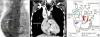
A 34-year-old man diagnosed with heterotaxy syndrome, unbalanced atrioventricular septal defect, double-outlet right ventricle, pulmonary artery (PA) stenosis, and totally anomalous pulmonary venous return was not a candidate for the Fontan procedure because of pulmonary hypertension (Supplementary Figure 1). However, he underwent PA banding at 29 years of age, and mean PA pressure decreased from 18 mmHg to 15 mmHg. Following bidirectional Glenn procedure and total abnormal pulmonary venous return repair performed in the subsequent year, cardiac dysfunction with atrioventricular valve regurgitation gradually developed with preserved right ventricular ejection fraction (RVEF). Therefore, extracardiac Fontan procedure using 24 mm expanded polytetrafluoroethylene graft with 5 mm-fenestration1) and common atrioventricular valve replacement were performed. Postsurgically, cyanosis and heart failure worsened. Decreased oxygen saturation (SpO2: 75% from 88%), elevated brain natriuretic peptide (BNP) levels (548 from 161 pg/mL) with NYHA class III, and high liver stiffness (26.6 ± 4.5 kPa) were observed. Cardiac angiography revealed reduced RVEF (29%) with ejection of venous blood into the systemic circulation through the fenestration (Figure 1) (Movie 1). Considering the risk of closing fenestration for elevated pressure to systemic venous pressure or decreased cardiac output, we intensified medical therapy for heart failure. Following β-blocker and angiotensin II receptor blocker administration, RVEF gradually improved (40%), BNP levels decreased (28 pg/mL), and NYHA class improved (stage II) with SpO2 83%. We planned using pulmonary vasodilators as an additional therapy when cyanosis and heart failure worsened.
Fontan fenestration leads to increased cardiac output and a concomitant decrease in venous congestion; however, it is detrimental to arterial saturation.2) The high PA pressure for a long time might be a risk factor for cyanosis and heart failure after surgery. For heart failure following a fenestrated Fontan procedure, medication could improve the function and pressure load of the RV.

 XML Download