

 PDF
PDF ePub
ePub Citation
 Print
Print


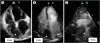
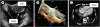
A 70-year-old female patient was transferred to our institution from a community hospital for coronary angiography after a non ST-elevation myocardial infarction. Her medical history was significant for the presence of Rosai Dorfman disease (RDD) for which was treated with low dose methylprednisolone. She had known abdominal lymphadenopathy and had also undergone right nephrectomy 5 years earlier due to right kidney involvement. At that time there was no cardiac involvement. Coronary angiography disclosed an intermediate severity stenosis of the right coronary artery and conservative management was decided. Transthoracic echocardiography revealed a sizeable mass of 4.5 × 4 cm in the right atrium showing enhancement after injection of echocardiographic contrast agent (Figure 1). Transesophageal echocardiography disclosed the presence of a right atrial mass encircling the cavity. It involved the right atrial free wall and the interatrial septum. It surrounded the ostium of the superior vena cava and the coronary sinus without causing obstruction (Figure 2). The mass was considered as the recurrence of past same disease and no other confirmative diagnostic process was made, regarding that there was no extra-nodal involvement. Due to the absence of hemodynamic abnormalities, we decided against interventional treatment and the patient was advised on periodic clinical and echocardiographic screening. NSTEMI could not be associated with RDD because the mass is not located near its right coronary artery orifice.
RDD is a very rare, non-malignant disease caused by proliferation of non-Langerhans sinus histiocytes.1) It is characterized of massive, painless and mainly bilateral cervical lymphadenopathy in childhood or early adulthood.2) Cardiac involvement is rare in RDD with only 17 cases previously reported in the English literature. Of these, the right atrium was affected in 7 while in some cases there was involvement in more than one cardiac locations.3) Treatment options include corticosteroids chemotherapy, radiotherapy and surgery local phenomena.2)
 XML Download