

 PDF
PDF ePub
ePub Citation
 Print
Print


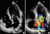
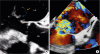
A 69-year-old male patient admitted to the hospital with congestive heart failure. Physical examination presented signs of left heart failure, and cardiac auscultation disclosed a grade III/VI pansystolic murmur at the apex. Transthoracic echocardiography showed a large saccular bulge originating from the anterior mitral valve leaflet with severe mitral regurgitation, and multiple valvular and subvalvular vegetations (Figure 1). Transesophageal echocardiography confirmed the diagnosis of complicated endocarditis of the mitral valve with large ruptured anterior mitral valve leaflet aneurysm (2.2 × 2.5 cm) producing severe mitral regurgitation (Figure 2). Aortic valve was clear of vegetations although it presented with mild to moderate aortic regurgitation. Blood cultures were positive for coagulase negative staphylococcus. A few days later, the patient underwent cardiac surgery for mitral and aortic valve replacement. The patient completed a 6-week antibiotic regimen following surgery and was then successfully discharged with no signs of heart failure.
Mitral valve aneurysms are rare and potentially fatal. Although they can occur in a non-infectious setting, most frequently they are a consequence of infective endocarditis and affect the anterior leaflet of the mitral valve.1) It is thought that infection causes leaflet degeneration through localized inflammation, dissection and consequent expansion of the valvular tissue towards the left atrium.2) Although conservative management with close follow-up may be possible in some aneurysms, mitral valve replacement surgery should be promptly considered in complicated cases.3)
This case highlights the importance of appropriate imaging for early detection of valvular complications and timely surgical intervention to achieve favorable outcomes.

 XML Download