Journal List > J Clin Neurol > v.15(4) > 1134815

Jung, Kim, Yu, O, Kim, Song, Kim, Kim, Heo, Park, Kim, Park, Choi, Park, Kim, Choi, Hwang, Chung, Bang, Kim, and Seo: Long-Term Outcomes of Real-World Korean Patients with Atrial-Fibrillation-Related Stroke and Severely Decreased Ejection Fraction

Abstract

Background and Purpose

The clinical implications of echocardiography findings for long-term outcomes in atrial fibrillation (AF)-related stroke patients are unknown.

Methods

This was a substudy of the Korean ATrial fibrillaTion EvaluatioN regisTry in Ischemic strOke patieNts (K-ATTENTION), which is a multicenter-based cohort comprising prospective stroke registries from 11 tertiary centers. Stroke survivors who underwent two-dimensional transthoracic echocardiography during hospitalization were enrolled. Echocardiography markers included the left-ventricle (LV) ejection fraction (LVEF), the left atrium diameter, and the ratio of the peak transmitral filling velocity to the mean mitral annular velocity during early diastole (E/e′ ratio). LVEF was categorized into normal (≥55%), mildly decreased (>40% and <55%), and severely decreased (≤40%). The E/e′ ratio associated with the LV filling pressure was categorized into normal (<8), borderline (≥8 and <15), and elevated (≥15). Kaplan-Meier and Cox regression analyses were performed for recurrent stroke, major adverse cardiac events, and all-cause death.

Results

This study finally included 1,947 patients. Over a median follow-up of 1.65 years (interquartile range, 0.42–2.87 years), the rates of recurrent stroke, major adverse cardiac events, and all-cause death were 35.1, 10.8, and 69.6 cases per 1,000 person-years, respectively. Multivariable analyses demonstrated that severely decreased LVEF was associated with a higher risks of major adverse cardiac events [hazard ratio (HR), 3.91; 95% confidence interval (CI), 1.58–9.69] and all-cause death (HR, 1.95; 95% CI, 1.23–3.10). The multivariable fractional polynomial plot indicated that recurrent stroke might be associated with a lower LVEF.

Conclusions

Severe LV systolic dysfunction could be a determinant of long-term outcomes in AF-related stroke.

INTRODUCTION

Atrial fibrillation (AF) is a well-recognized risk factor for stroke and mortality, and it also confers a higher risk of coronary artery diseases, including myocardial infarction (MI).1 The coexistence of AF in high-risk patients such as those with ischemic stroke (so-called AF-related stroke) markedly increases the risk of thromboembolic events and mortality compared to those with a normal sinus rhythm.234 However, the influence of AF on long-term outcomes of ischemic stroke remains unclear because these findings are mostly based on observational studies performed prior to the era of direct oral anticoagulants (DOACs). Few studies have investigated the long-term prognostic implications of AF with ischemic stroke since the introduction of DOACs, which prevail nowadays.5 The prescribing of DOACs for the purpose of stroke prevention has increased markedly in South Korea since 2013,6 which makes it essential to identify outcome-related factors in AF-related stroke by performing long-term clinical observations following the introduction of DOACs in order to reduce the future risk of vascular events and mortality in these patients.
Transthoracic echocardiography (TTE) has become the routine stroke workup for identifying embolic sources or comorbidities of the cardiopulmonary system. It was recently reported that the left-ventricle (LV) ejection fraction (LVEF) as measured in TTE could provide long-term prognostic information for predicting the risk of stroke and mortality in AF7 as well as ischemic stroke patients.89 However, these studies have produced conflicting results, probably due to heterogeneity in the study design, patient cohort, race, AF type, anticoagulation treatment, variable diagnostic criteria for systolic LV dysfunction, and small samples. Additionally, the clinical implications of LVEF remain unknown in high-risk individuals with concomitant AF and ischemic stroke.
Our aim was to determine the prognostic value of TTE features including LVEF for long-term outcomes including recurrent stroke, major adverse cardiac events, and all-cause death in AF-related stroke.

METHODS

Study design and participants

This study was a substudy of the Korean ATrial fibrillaTion EvaluatioN regisTry in Ischemic strOke patieNts (K-ATTENTION), which is a real-world cohort comprising prospective stroke registries from 11 tertiary centers of South Korea being used to investigate the characteristics, use of oral anticoagulants, and outcomes in AF-related stroke patients. Among ischemic stroke patients who were admitted within 7 days of stroke onset between January 2013 and December 2015, those with AF-related stroke regardless of its subtype (lacunar, atherothrombotic, or cardioembolic) were consecutively enrolled. This study finally included only stroke survivors who underwent two-dimensional TTE during hospitalization in order to evaluate the effects of echocardiography findings on long-term outcomes. Stroke neurologists in each hospital were in charge of caring for the stroke patients during hospitalization and follow-up. They usually followed the usual stroke guidelines regarding the evaluation, acute treatment, and acute and long-term prevention of stroke. Additionally, the type or dosage of antithrombotic agents at discharge was decided while considering the functional status, individual patient preference, complications such as hemorrhagic transformation, and compliance.
This study was approved by the Institutional Review Board of each center. The IRB number of the affiliated center of the first author was 2016AS0051. Informed consent was not required due to the retrospective design of the study.

Basic evaluation

The detailed clinical information included the National Institutes of Health Stroke Scale (NIHSS) score, modified Rankin Scale (mRS) score, potential risk factors, and management during admission and at discharge. Potential risk factors included sex, smoking status, congestive heart failure (CHF), hypertension, diabetes mellitus, coronary artery disease, peripheral artery disease, and prior stroke or transient ischemic attack. The CHADS2 and CHA2DS2-VASc scores, which account for risk factors including age, sex, hypertension, diabetes mellitus, CHF, and vascular diseases, of each patient were estimated based on the time before the index stroke.
Ischemic lesions were confirmed by diffusion-weighted MRI or CT. The vascular status was evaluated using MRI or CT angiography, if needed, or cerebral angiography. Laboratory examinations included the complete blood count and the total cholesterol, triglyceride, high-density cholesterol, low-density cholesterol, and creatinine levels. Chronic kidney disease (CKD) was defined as an estimated glomerular filtration rate of <60 mL/min/1.73 m2 using the CKD Epidemiology Collaboration creatinine equation. Twelve-lead electrocardiography (ECG) was performed at admission and repeated when necessary. AF was documented on ECG during hospitalization and/or 24-hour Holter monitoring and/or continuous ECG monitoring in the stroke unit. AF was classified into paroxysmal vs. sustained5 and nonvalvular vs. valvular. Valvular AF was defined as cases accompanied by a moderate degree of mitral stenosis or mechanical valve replacement.

Echocardiography evaluation and parameters

TTE was performed as soon as possible after admission, and the findings were interpreted by a cardiologist in each center. Because TTE protocols can differ among centers, we extracted common parameters when performing TTE including the left atrium (LA) diameter (LAD), LVEF, and the ratio of the peak transmitral filling velocity to the mean mitral annular velocity during early diastole (E/e′ ratio). LAD was measured as the maximum anteroposterior diameter of the LA in the parasternal long-axis view at end systole.10 Due to sex-based differences in LAD, indexed LAD (iLAD) was calculated by dividing LAD (cm) by the body surface area (m2).10 The iLAD values were categorized into quartiles. LVEF was calculated from estimates of the end-systolic and end-diastolic LV volumes using the modified Simpson's rule. LVEF was categorized into normal (≥55%), mildly decreased (>40% and <55%), and severely decreased (≤40%) based on research evaluating LVEF as the outcome determinant in acute ischemic stroke.11 The peak transmitral filling velocity (E) was measured using the pulsed-wave Doppler method. The mean mitral annular velocity at early diastole (e′) was measured using the tissue Doppler method from the septal corner of the mitral annulus in the apical four-chamber view. The E/e′ ratio associated with the LV filling pressure was categorized into normal (<8), borderline (≥8 and <15), and elevated (≥15).

Outcomes of interest and follow-up

Recurrent stroke, major adverse cardiac events, and allcause death were the outcomes of interest. In particular, recurrent stroke implies ischemic stroke and major adverse cardiac events consisting of acute MI, unstable angina, or coronary revascularization procedures including a percutaneous coronary intervention and coronary artery bypass graft.
The follow-up was performed up to January 2018. Follow-up data were acquired through medical record reviews at outpatient clinics. For a patient who was unable to complete clinic visits, a telephone interview was performed by an experienced research nurse at each center to obtain the required information.

Statistical analyses

If there were any missing values regarding laboratory findings, provided the prevalence of that variable was <3%, the mean value for those variables was used in order to reduce the loss of patient information. Categorical variables are presented as number (percentage) values, and continuous variables are presented as mean±SD deviation or median (interquartile range, IQR) values, as appropriate. We used the Kolmogorov-Smirnov test to evaluate whether continuous variables conformed to a normal distribution. A simple comparison of LVEF grade was performed using the χ2 test for categorical variables and one-way ANOVA or the Wilcoxon signed-rank test for continuous variables depending on conformity with a normal distribution. To compare cumulative event rates between groups using the Kaplan-Meier method, the Breslow or log-rank test was applied depending on whether or not the survival graphs intersected. Multivariable Cox regression analyses were used to identify significant predictors of specific outcomes.
All variables in univariable analyses for which p<0.1 as well as LVEF as an important echocardiography marker were included in the final models of specific outcomes. Multicollinearity was assessed by assessing the variance inflation factor, especially that between echocardiography markers. Sensitivity analyses were performed by imputing LVEF as a continuous variable or according to another LVEF criterion9 [normal (>35%) vs. low (≤35%)] to confirm the robustness of the effect of LVEF on long-term outcomes. Multivariable fractional polynomial plots were used to investigate the effect of LVEF on each of the outcomes. Additional models were developed in which CHADS2 or CHA2DS2-VASc scores were imputed rather than using relevant clinical covariates.
We performed subgroup analyses of outcomes that were significantly related to LVEF. An a-priori subgroup analysis was applied to age (≥75 and <75 years old), sex, body mass index (BMI) (underweight, <18.5 kg/m2; normal weight, 18.5–22.9 kg/m2; overweight, 23.0–24.9 kg/m2; obese, ≥25 kg/m2), initial NIHSS score (mild, 0–4; moderate, 5–15; severe, 16–20; very severe, ≥21), thrombolysis type, AF type, iLAD, LV filling pressure, discharge medication including antithrombotic agents and statins, and discharge mRS score (0–3 and 4 or 5) by including each of the interaction terms in specific Cox multivariable models.
All analyses were performed using SPSS software (version 20.0 for Windows, IBM Corp., Armonk, NY, USA), MedCalc software (version 16.4.1, Mariakerke, Belgium), and STATA software (version 13, StataCorp, College Station, TX, USA)

RESULTS

Baseline characteristics

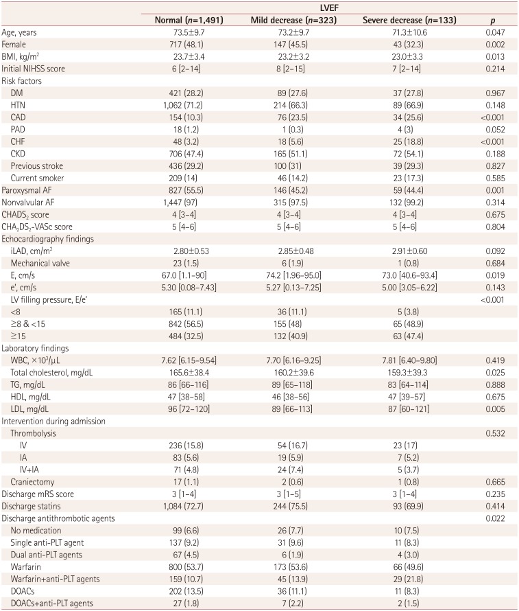
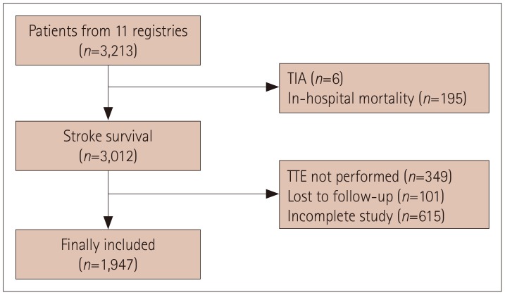
Among the 3,213 included patients, 3,012 survived their index stroke after admission. Compared to the excluded patients, the included ones had lower prevalence rates of being female and having CHF and a previous stroke history, were associated with less-severe stroke at admission and discharge, and had statins and DOACs prescribed more frequently to them at discharge (Supplementary Table 1 in the online-only Data Supplement).
The study finally included 1,947 patients (Fig. 1) aged 73.3±9.8 years, with 1,040 (53.4%) males. Nonvalvular AF was present in 1,894 patients, while valvular AF was observed in 54 (2.8%) patients: 25 with moderate-to-severe mitral stenosis, and 29 with mechanical valve replacement. On the other hand, 1,032 (53%) patients had paroxysmal AF, whereas sustained AF was classified in 915 (47%). The median CHADS2 and CHA2DS2-VASc scores were 3 (IQR, 3–4) and 5 (IQR, 4–6), respectively. Discharge medications of single antiplatelet agents, dual antiplatelet agents, warfarin, warfarin+antiplatelet agents, DOACs, and DOACs+antiplatelet agents were prescribed to 179 (9.2%), 77 (4.0%), 1,039 (53.4%), 233 (12.0%), 249 (12.8%), and 36 (1.8%) patients, respectively. Dabigatran was the most frequently prescribed DOACs (n=99, 39.8%), followed by apixaban (n=80, 32.1%) and rivaroxaban (n=72, 28.9%). Edoxaban was not prescribed prior to its official release in South Korea. No antithrombotic medications were prescribed at discharge to 134 (6.9%) patients.

Comparison by LVEF grade

Patients were classified into three groups according to LVEF grade (Table 1). The initial stroke severity, thrombolysis type, functional status at discharge, and statins prescribed at discharge were similar among the three groups. A higher usage of warfarin+antiplatelet agents and a lower use of DOACs at discharge was observed in the severely decreased LVEF group, probably due to the prevalence of comorbid coronary artery disease being higher than in the other groups. Severely decreased LVEF was associated with male sex, younger age, and lower BMI. Patients in this group frequently exhibited cardiac diseases including CHF, coronary artery disease, and sustained AF. Among the echocardiography markers, the E/e′ ratio was associated with the LVEF grade.

Outcomes and echocardiography markers

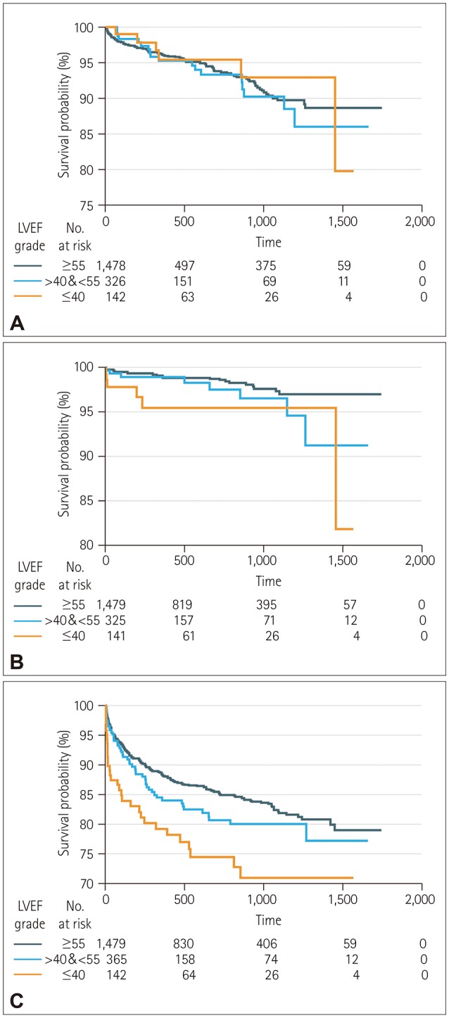
Over a median follow-up of 1.65 years (IQR, 0.42–2.87 years), 113 patients experienced recurrent stroke, 36 experienced major adverse cardiac events, and 236 died. The rates of recurrent stroke, major adverse cardiac events, and all-cause death were 35.1, 10.8, and 69.6 cases per 1,000 person-years, respectively. Kaplan-Meyer curves demonstrated that the cumulative probability of major adverse cardiac events (Breslow test, p=0.005) and all-cause death (Breslow test, p=0.032), but not recurrent stroke, was higher in patients in the severely decreased LVEF group than in the other groups (Fig. 2). Other echocardiography parameters were not related to any other outcomes of interest.

Multivariable analyses

Table 2 summarizes the results obtained in the multivariable analyses. No echocardiography features were associated with recurrent stroke. In contrast, a severely decreased LVEF was associated with an increased risk of major adverse cardiac events and all-cause death compared to the normal-LVEF group. After inputting LVEF as a continuous variable or another LVEF criterion, the increased risk of lower LVEF persisted for major adverse cardiac events and all-cause death. The multivariable fractional polynomial plots in Fig. 3 indicate that a lower LVEF was associated with higher risks of recurrent stroke, major adverse cardiac events, and all-cause death. In terms of recurrent stroke, a lower LVEF (particularly <50%) was associated with a higher risk of recurrent stroke, although a severely decreased LVEF was not correlated with recurrent stroke in the multivariable Cox model.
After adjusting for the CHADS2 or CHA2DS2-VASc score, no association with any echocardiography feature was found, although these risk stratification tools were reported as predictors for recurrent stroke in this study (adjusted hazard ratio, 1.36 per 1-point increase for the CHADS2 score, and 1.23 per 1-point increase for the CHA2DS2-VASc score; Supplementary Tables 2 and 3 in the online-only Data Supplement). In contrast, a severely decreased LVEF was associated with increased risks of major adverse cardiac events and all-cause death compared to the normal LVEF group. Considering the CHA2DS2-VASc score, a severely decreased LVEF could marginally predict an increased risk of all-cause death.

Subgroup analyses and interactions

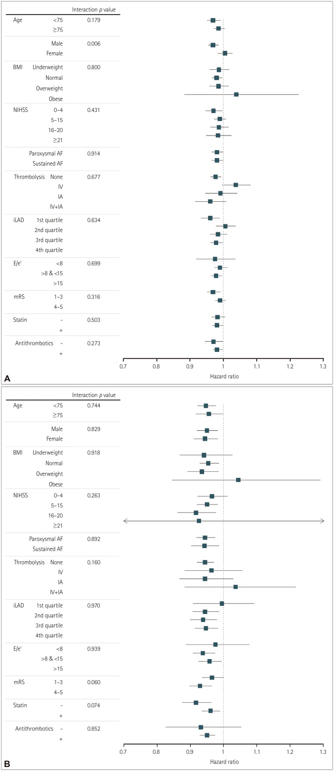
Subgroup analyses were performed of all-cause death and major adverse cardiac events (Fig. 4). The effect of a lower LVEF on increasing the risk of all-cause death and major adverse cardiac events was comparatively consistent across prespecified subgroups, with no significant interaction except for sex in all-cause death. The prognostic role of LVEF in all-cause death was lower for female sex than for male sex.
For recurrent stroke, a significant interaction was found between the severely decreased LVEF group and LVEF as a continuous variable (p=0.048), suggesting that a lower LVEF is associated with recurrent stroke.

DISCUSSION

This study is the first to demonstrate a prognostic effect of global LV systolic dysfunction (measured as LVEF) in acute ischemic stroke patients with AF. We focused on stroke survivors in order to scrutinize the long-term effects of LVEF in AF-related stroke and such effects according to various types of AF. In addition, this study substantially reflected the real-world clinical practice of stroke neurologists at each center around the transition time after the introduction of DOACs in South Korea.
Recent studies have demonstrated that the mortality rate of AF patients has remained high despite the risk of thromboembolic events and mortality having decreased through the use of oral anticoagulants.1213 This study found that all-cause death occurred 1.78 times more frequently than recurrent stroke during the 1.65 years of follow-up, suggesting that death could be still a major health issue in these patients. Additionally, severe LV systolic dysfunction as detected using two-dimensional TTE was revealed as a meaningful predictor of all-cause death. Although heart failure (HF) is reportedly an important prognostic factor for mortality in patients with AF,1415 we found that LVEF rather than the presence of HF to play the important role. This could mean that the presence of HF could be underestimated in admissions to neurology departments, because HF is a clinical syndrome with typical symptoms and signs, and additionally is associated with abnormal plasma natriuretic peptide levels and echocardiography findings.16 This finding remained even after performing adjustments using risk stratification tools, including the CHADS2 and CHA2DS2-VASc scores that have recently been reported as prognostic markers.1718 Also, the association between LVEF and all-cause death was found regardless of the prespecified LVEF criteria of LV dysfunction. This is consistent with two studies demonstrating an association between LVEF as measured using TTE and long-term mortality in ischemic stroke.89 However, those studies included all ischemic stroke patients regardless of the presence of AF. A small study that included only 132 stroke patients without AF found no impact of LV systolic dysfunction on long-term mortality.19 On the other hand, LV systolic dysfunction was reportedly associated with mortality in patients with AF.20 Together these previous findings and the present results suggest that LV systolic dysfunction per se is an important outcome predictor for mortality in ischemic stroke patients, especially in the presence of AF.
All of the present subgroup analyses revealed a similar pattern and direction of the correlation between LVEF and all-cause death, but not for sex. Males with a low LVEF tended to have an increased risk of mortality. This sex-based difference may be attributable to a discrepancy between differences in baseline characteristics, medications at discharge, or genetic inconsistencies. However, the exact cause of this difference was not identified in the present study. Meanwhile, a severely decreased LVEF conferred a high risk of major adverse cardiac events. These findings were consistent in all of the subgroup analyses.
Like risk stratification schemes including the CHADS2 or CHA2DS2-VASc score, LVEF might predict recurrent stroke. However, a possible association with lower LVEF (particularly <40%) was found only in the multivariable fractional plot and interaction analysis. This means that care is needed during interpretations, which is due to a low statistical power associated with the small number of recurrent strokes that occurred in this lower LVEF group. A reduced LVEF is known to be a risk factor for stroke, most often in MI survivors or CHF patients.21 With regard to the secondary prevention of ischemic stroke, the effect of LVEF is limited in that the criterion of a very severely decreased LVEF (<15%) as a predictor was applied to patients with CHF.22 However, the predictive role of LV systolic dysfunction based on echocardiography for stroke has been inconsistent in AF patients.23 In addition, in patients with concomitant ischemic stroke and AF, the association between low LVEF and recurrent stroke remains unknown due to a lack of relevant studies, although some studies have found the size or diameter of the LA to be a predictor for stroke besides AF.242526 Care is required with interpreting the LAD due to sex-based variations.27 Compared to previous positive results, our study demonstrated that the sex-adjusted LAD in terms of secondary prevention for stroke in AF-related stroke patients was not related to recurrent stroke. This discrepancy might be attributable to differences among the included patients (e.g., mean age and sex proportions) and in the inclusion criteria (nonvalvular AF vs. all-type AF). Other echocardiography markers of cardiac function and structure such as LV geometry, global longitudinal strain,28 and LA appendage wall velocity29 as measured using transesophageal echocardiography (TEE) have recently be proposed as risk factors. Further studies that needed to comprehensively evaluate echocardiography markers obtained using TTE and/or TEE (if possible) as predictors for recurrent stroke in AF-related stroke.
This study was subject to several limitations. First, the use of a prospective registry is susceptible to selection bias as well as unmeasured bias that is inherent in observational studies. In the present study, several characteristics of the excluded patients in whom TTE was not performed differed from those of the included patients. Second, LVEF has limitations including geometric assumptions, load dependency, reproducibility, interobserver variability, and the influence of heart rate (e.g., AF) and translational motion, although it is the most widely used, convenient, and significant parameter for patient classification and treatment decisions.30 Third, this study did not consider or adjust for information regarding the maintenance, change, or withdrawal of anticoagulants, although maintaining the administration of an anticoagulant is known to lower the risk of thrombotic events. In addition, the effect of DOAC usage might not have been reflected in the outcomes due to the transition time after its introduction.
In conclusion, severe LV systolic dysfunction could be a determinant of long-term outcomes including all-cause death and major adverse cardiac events in patients with AF-related stroke. In particular, recurrent stroke might develop in patients with severe LV dysfunction. LV systolic function measured as the LVEF could help clinicians in the stroke field to identify patients at high risks of all-cause death, major adverse cardiac events, and recurrent stroke. In addition, the present results emphasize their need to pay attention to and manage LV systolic function beyond anticoagulation in order to further reduce these long-term outcomes in AF-related stroke patients.

Acknowledgements

This study was supported by a grant from the Korean Neurological Association (KNA-17-MI-10).
The authors appreciate Jaehyung Cha, statistician from Medical Science Research Center, Korea University Ansan Hospital, for his help with the data analysis.

Notes

Author Contributions:

  • Conceptualization: Jin-Man Jung, Yong-Hyun Kim, Woo-Keun Seo.

  • Data curation: Jin-Man Jung, Sungwook Yu, Kyungmi O, Chi Kyung Kim, Tae-Jin Song, Yong-Jae Kim, Bum Joon Kim, Sung Hyuk Heo, Kwang-Yeol Park, Jeong-Min Kim, Jong-Ho Park, Jay Chol Choi, Man-Seok Park, Joon-Tae Kim, Kang-Ho Choi, Yang-Ha Hwang, Jong-Won Chung, Oh Young Bang, Gyeong-moon Kim.

  • Formal analysis: Jin-Man Jung, Woo-Keun Seo.

  • Funding acquisition: Woo-Keun Seo.

  • Investigation: Jin-Man Jung, Sungwook Yu, Kyungmi O, Chi Kyung Kim, Tae-Jin Song, Yong-Jae Kim, Bum Joon Kim, Sung Hyuk Heo, Kwang-Yeol Park, Jeong-Min Kim, Jong-Ho Park, Jay Chol Choi, Man-Seok Park, Joon-Tae Kim, Kang-Ho Choi, Yang-Ha Hwang, Jong-Won Chung, Oh Young Bang, Gyeong-moon Kim.

  • Methodology: Jin-Man Jung, Yong-Hyun Kim, Woo-Keun Seo.

  • Project administration: Woo-Keun Seo.

  • Resources: Jin-Man Jung, Yong-Hyun Kim, Woo-Keun Seo.

  • Software: Jin-Man Jung, Woo-Keun Seo.

  • Supervision: Woo-Keun Seo.

  • Validation: Jin-Man Jung, Woo-Keun Seo.

  • Visualization: Jin-Man Jung, Woo-Keun Seo.

  • Writing—original draft: Jin-Man Jung, Woo-Keun Seo.

  • Writing—review & editing: Sungwook Yu, Kyungmi O, Chi Kyung Kim, Tae-Jin Song, Yong-Jae Kim, Bum Joon Kim, Sung Hyuk Heo, Kwang-Yeol Park, Jeong-Min Kim, Jong-Ho Park, Jay Chol Choi, Man-Seok Park, Joon-Tae Kim, Kang-Ho Choi, Yang-Ha Hwang, Jong-Won Chung, Oh Young Bang, Gyeong-moon Kim, Woo-Keun Seo.

Conflicts of Interest: The authors have no potential conflicts of interest to disclose.

References

1. Guo XY, Li N, Du X, Bai R, Yu RH, Long DY, et al. Atrial fibrillation is associated with an increased risk of myocardial infarction: insights from a meta-analysis. Atherosclerosis. 2016; 254:1–7. PMID: 27673698.
crossref
2. Kaarisalo MM, Immonen-Räihä P, Marttila RJ, Salomaa V, Kaarsalo E, Salmi K, et al. Atrial fibrillation and stroke. Mortality and causes of death after the first acute ischemic stroke. Stroke. 1997; 28:311–315. PMID: 9040681.
3. Marini C, De Santis F, Sacco S, Russo T, Olivieri L, Totaro R, et al. Contribution of atrial fibrillation to incidence and outcome of ischemic stroke: results from a population-based study. Stroke. 2005; 36:1115–1119. PMID: 15879330.
4. Lin HJ, Wolf PA, Kelly-Hayes M, Beiser AS, Kase CS, Benjamin EJ, et al. Stroke severity in atrial fibrillation. The Framingham Study. Stroke. 1996; 27:1760–1764. PMID: 8841325.
5. Koga M, Yoshimura S, Hasegawa Y, Shibuya S, Ito Y, Matsuoka H, et al. Higher risk of ischemic events in secondary prevention for patients with persistent than those with paroxysmal atrial fibrillation. Stroke. 2016; 47:2582–2588. PMID: 27531346.
crossref
6. Lee SR, Choi EK, Han KD, Cha MJ, Oh S, Lip GYH. Temporal trends of antithrombotic therapy for stroke prevention in Korean patients with non-valvular atrial fibrillation in the era of non-vitamin K antagonist oral anticoagulants: a nationwide population-based study. PLoS One. 2017; 12:e0189495. PMID: 29261716.
crossref
7. Simpson E, Stevenson M, Scope A, Poku E, Minton J, Evans P. Echocardiography in newly diagnosed atrial fibrillation patients: a systematic review and economic evaluation. Health Technol Assess. 2013; 17:1–263. v–vi.
crossref
8. Miles JA, Garber L, Ghosh S, Spevack DM. Association of transthoracic echocardiography findings and long-term outcomes in patients undergoing workup of stroke. J Stroke Cerebrovasc Dis. 2018; 27:2943–2950. PMID: 30072178.
crossref
9. Milionis H, Faouzi M, Cordier M, D'Ambrogio-Remillard S, Eskandari A, Michel P. Characteristics and early and long-term outcome in patients with acute ischemic stroke and low ejection fraction. Int J Cardiol. 2013; 168:1082–1087. PMID: 23176765.
crossref
10. Lang RM, Badano LP, Mor-Avi V, Afilalo J, Armstrong A, Ernande L, et al. Recommendations for cardiac chamber quantification by echocardiography in adults: an update from the American Society of Echocardiography and the European Association of Cardiovascular Imaging. Eur Heart J Cardiovasc Imaging. 2015; 16:233–270. PMID: 25712077.
crossref
11. Kim WJ, Nah HW, Kim DH, Cha JK. Association between left ventricular dysfunction and functional outcomes at three months in acute ischemic stroke. J Stroke Cerebrovasc Dis. 2016; 25:2247–2252. PMID: 27449114.
crossref
12. Gupta DK, Giugliano RP, Ruff CT, Claggett B, Murphy S, Antman E, et al. The prognostic significance of cardiac structure and function in atrial fibrillation: the ENGAGE AF-TIMI 48 echocardiographic substudy. J Am Soc Echocardiogr. 2016; 29:537–544. PMID: 27106009.
13. Friberg L, Hammar N, Pettersson H, Rosenqvist M. Increased mortality in paroxysmal atrial fibrillation: report from the Stockholm Cohort-Study of Atrial Fibrillation (SCAF). Eur Heart J. 2007; 28:2346–2353. PMID: 17670754.
crossref
14. Wang TJ, Larson MG, Levy D, Vasan RS, Leip EP, Wolf PA, et al. Temporal relations of atrial fibrillation and congestive heart failure and their joint influence on mortality: the Framingham Heart Study. Circulation. 2003; 107:2920–2925. PMID: 12771006.
15. Marijon E, Le Heuzey JY, Connolly S, Yang S, Pogue J, Brueckmann M, et al. Causes of death and influencing factors in patients with atrial fibrillation: a competing-risk analysis from the randomized evaluation of long-term anticoagulant therapy study. Circulation. 2013; 128:2192–2201. PMID: 24016454.
16. Kim W, Kim EJ. Heart failure as a risk factor for stroke. J Stroke. 2018; 20:33–45. PMID: 29402070.
crossref
17. Jover E, Roldán V, Gallego P, Hernández-Romero D, Valdés M, Vicente V, et al. Predictive value of the CHA2DS2-VASc score in atrial fibrillation patients at high risk for stroke despite oral anticoagulation. Rev Esp Cardiol (Engl Ed). 2012; 65:627–633. PMID: 22609214.
18. Tanaka K, Yamada T, Torii T, Furuta K, Matsumoto S, Yoshimura T, et al. Pre-admission CHADS2, CHA2DS2-VASc, and R2CHADS2 scores on severity and functional outcome in acute ischemic stroke with atrial fibrillation. J Stroke Cerebrovasc Dis. 2015; 24:1629–1635. PMID: 25906940.
19. Holmström A, Fu ML, Hjalmarsson C, Bokemark L, Andersson B. Heart dysfunction in patients with acute ischemic stroke or TIA does not predict all-cause mortality at long-term follow-up. BMC Neurol. 2013; 13:122. PMID: 24053888.
crossref
20. Stöllberger C, Chnupa P, Abzieher C, Länger T, Finsterer J, Klem I, et al. Mortality and rate of stroke or embolism in atrial fibrillation during long-term follow-up in the embolism in left atrial thrombi (ELAT) study. Clin Cardiol. 2004; 27:40–46. PMID: 14743856.
crossref
21. Ferreira JP, Girerd N, Gregson J, Länger T, Finsterer J, Klem I, et al. Stroke risk in patients with reduced ejection fraction after myocardial infarction without atrial fibrillation. J Am Coll Cardiol. 2018; 71:727–735. PMID: 29447733.
crossref
22. Pullicino PM, Qian M, Sacco RL, Freudenberger R, Graham S, Teerlink JR, et al. Recurrent stroke in the warfarin versus aspirin in reduced cardiac ejection fraction (WARCEF) trial. Cerebrovasc Dis. 2014; 38:176–181. PMID: 25300706.
crossref
23. Agarwal M, Apostolakis S, Lane DA, Lip GY. The impact of heart failure and left ventricular dysfunction in predicting stroke, thromboembolism, and mortality in atrial fibrillation patients: a systematic review. Clin Ther. 2014; 36:1135–1144. PMID: 25146364.
crossref
24. Yaghi S, Moon YP, Mora-McLaughlin C, Willey JZ, Cheung K, Di Tullio MR, et al. Left atrial enlargement and stroke recurrence: the Northern Manhattan Stroke Study. Stroke. 2015; 46:1488–1493. PMID: 25908460.
crossref
25. Broughton ST, O'Neal WT, Salahuddin T, Soliman EZ. The influence of left atrial enlargement on the relationship between atrial fibrillation and stroke. J Stroke Cerebrovasc Dis. 2016; 25:1396–1402. PMID: 27012217.
crossref
26. Paciaroni M, Agnelli G, Falocci N, Caso V, Becattini C, Marcheselli S, et al. Prognostic value of trans-thoracic echocardiography in patients with acute stroke and atrial fibrillation: findings from the RAF study. J Neurol. 2016; 263:231–237. PMID: 26566907.
crossref
27. Ogata T, Matsuo R, Kiyuna F, Hata J, Ago T, Tsuboi Y, et al. Left atrial size and long-term risk of recurrent stroke after acute ischemic stroke in patients with nonvalvular atrial fibrillation. J Am Heart Assoc. 2017; 6:e006402. PMID: 28862939.
crossref
28. Kalam K, Otahal P, Marwick TH. Prognostic implications of global LV dysfunction: a systematic review and meta-analysis of global longitudinal strain and ejection fraction. Heart. 2014; 100:1673–1680. PMID: 24860005.
crossref
29. Tamura H, Watanabe T, Nishiyama S, Sasaki S, Wanezaki M, Arimoto T, et al. Prognostic value of low left atrial appendage wall velocity in patients with ischemic stroke and atrial fibrillation. J Am Soc Echocardiogr. 2012; 25:576–583. PMID: 22326133.
crossref
30. Nesbitt GC, Mankad S, Oh JK. Strain imaging in echocardiography: methods and clinical applications. Int J Cardiovasc Imaging. 2009; 25 Suppl 1:9–22. PMID: 19145475.
crossref

Supplementary Materials

The online-only Data Supplement is available with this article at https://doi.org/10.3988/jcn.2019.15.4.545.

Supplementary Table 1

Comparison of included and excluded stroke survivors
jcn-15-545-s001.pdf

Supplementary Table 2

Multivariable analysis adjusted for CHADS2 score
jcn-15-545-s002.pdf

Supplementary Table 3

Multivariable analysis adjusted for CHA2DS2-VASc score
jcn-15-545-s003.pdf
Fig. 1

Flow chart of included patients. TIA: transient ischemic attack, TTE: transthoracic echocardiography.

jcn-15-545-g001
Fig. 2

Survival curves of recurrent stroke (A), major adverse cardiac events (B), and all-cause death (C). LVEF: left-ventricle ejection fraction.

jcn-15-545-g002
Fig. 3

Multivariable fractional polynomial plots. The x-axis shows the HR and the y-axis is the LVEF (%). A: Recurrent stroke. B: Major adverse cardiac events. C: All-cause death. HR: hazard ratio, LVEF: left-ventricle ejection fraction.

jcn-15-545-g003
Fig. 4

Results from subgroup analyses of all-cause death (A) and major adverse cardiac events (B). AF: atrial fibrillation, BMI: body mass index, E: peak transmitral filling velocity, e′: mean mitral annular velocity at early diastole, IA: intra-arterial, iLAD: indexed left atrium diameter, IV: intravenous, mRS: modified Rankin Scale, NIHSS: National Institutes of Health Stroke Scale.

jcn-15-545-g004
Table 1

Baseline characteristics of the included patients

jcn-15-545-i001

Data are mean±SD, n (%), or median [interquartile range] values. Missing values for the creatinine level (n=48, 2.46%) were substituted with the mean creatinine level.

AF: atrial fibrillation, BMI: body mass index, CAD: coronary artery disease, CHF: congestive heart failure, CKD: chronic kidney disease, DM: diabetes mellitus, DOACs: direct oral anticoagulants, E: peak transmitral filling velocity, e′: mean mitral annular velocity at early diastole, HDL: high-density lipoprotein, HTN: hypertension, IA: intra-arterial, iLAD: indexed left atrium diameter, IV: intravenous, LDL: lowdensity lipoprotein, LV: left-ventricle, LVEF: left-ventricle ejection fraction, mRS: modified Rankin Scale, NIHSS: National Institutes of Health Stroke Scale, PAD: peripheral artery disease, PLT: platelet, TG: triglyceride, WBC: white blood cells.

Table 2

Results from multivariable analyses of long-term outcomes

jcn-15-545-i002

Data are median [interquartile range] values. Cox multivariable models for recurrent stroke included sex, age, CHF, DM, AF type, and iLAD. Multivariable analyses of major adverse cardiac events included age, discharge mRS score, CHF, CAD, AF type, iLAD, CKD, and discharge medication. Multivariable analyses of all-cause death included sex, age, BMI, discharge mRS score, recurrent stroke, DM, iLAD, LV filling pressure, statin at discharge, and discharge medication. Multicollinearity was absent because all of the variance inflation factor levels were <2. Discharge medication was categorized into no medication, single antiplatelet agent, dual antiplatelet agents, warfarin, warfarin+antiplatelet agents, DOACs, and DOACs+antiplatelet agents.

*LVEF was used as a continuous rather than a categorical variable, LVEF was classified into normal (>35%) and low (≤35%).

AF: atrial fibrillation, BMI: body mass index, CAD: coronary artery disease, CHF: congestive heart failure, CI: confidence interval, CKD: chronic kidney disease, DM: diabetes mellitus, DOACs: direct oral anticoagulants, HR: hazard ratio, iLAD: indexed left atrium diameter, LV: left-ventricle, LVEF: left-ventricle ejection fraction, mRS: modified Rankin Scale.

TOOLS
Similar articles