Journal List > J Clin Neurol > v.15(4) > 1134810

Kim, Chung, Park, and Kim: Language-Related White-Matter-Tract Deficits in Children with Benign Epilepsy with Centrotemporal Spikes: A Retrospective Study

Abstract

Background and Purpose

Benign epilepsy with centrotemporal spikes (BECTS) is one of the most common pediatric epilepsies, and it generally has a good prognosis. However, recent research has indicated that the epileptic activity of BECTS can cause cognitive defects such as language, visuospatial, and auditory verbal memory deficits. This study assessed language-delivery deficits in BECTS patients using diffusion-tensor magnetic resonance imaging (DTI).

Methods

T1-weighted MRI, DTI, and language tests were conducted in 16 BECTS patients and 16 age-matched controls. DTI data were analyzed using the TRActs Constrained by Underlying Anatomy tool in FreeSurfer 5.3, and 18 major white-matter tracts were extracted, which included 4 language-related tracts: the inferior longitudinal fasciculus, superior longitudinal fasciculus-parietal terminations, superior longitudinal fasciculus-temporal terminations, and uncinate fasciculus (UNC). Language tests included the Korean version of the Receptive and Expressive Vocabulary Test, Test of Problem-Solving Abilities (TOPS), and the mean length of utterance in words.

Results

The BECTS group exhibited decreased mean fractional anisotropy and increased mean radial diffusivity, with significant differences in both the superior longitudinal fasciculus and the left UNC (p<0.05), which are the language-related white-matter tracts in the dual-loop model. The TOPS language test scores were significantly lower in the BECTS group than in the control group (p<0.05).

Conclusions

It appears that BECTS patients can exhibit language deficits. Seizure activities of BECTS could alter DTI scalar values in the language-related white-matter tracts.

INTRODUCTION

Benign epilepsy with centrotemporal spikes (BECTS) is one of the most common pediatric epilepsies and is well known to generally have a good prognosis. However, recent research has indicated that the epileptic activity of BECTS causes cognitive problems such as visuospatial and auditory verbal memory deficits,1 and especially language dysfunction.2 Various fundamental mechanisms have been proposed for the language dysfunction in BECTS.3 The dual-loop model reported in 1994 is the most prominent contemporary model of the language-delivery process, and it comprises ventral and dorsal streams that are divided around the Sylvian fissure.4 The dorsal and ventral streams involve phonological and semantic processing of language, respectively.5 The dorsal stream comprises the superior longitudinal fasciculus (SLF) and arcuate fasciculus (AF), and the ventral stream comprises the extreme capsule and the uncinate fasciculus (UNC), middle longitudinal fasciculus, inferior longitudinal fasciculus (ILF), and inferior fronto-occipital fasciculus.
Diffusion-tensor magnetic resonance imaging (DTI) can be employed to study neural fiber tracts in human subjects,6 and has been used to evaluate pediatric psychiatric diseases such as autism and attention deficit hyperactivity disorder (ADHD).78 The white-matter fibers associated with language have recently been studied using DTI.9
Neuroimaging studies using MRI have been performed for BECTS. Cognitive and behavioral deficits in BECTS are associated with the decreased volume of the frontal lobes.2 Furthermore, recent DTI studies of epilepsy have detected white-matter deficits.10 DTI showed a decrease in fractional anisotropy (FA) values—one of the DTI scalar values—at the focus of seizures in BECTS.11 DTI data can be analyzed using two methods: tract-based analysis and voxel-based analysis. Voxel-based analysis assesses an image of the white matter based on voxel stepwise locations, whereas tract-based analysis extracts the region of interest from the seed to the target-region tract.12 Although voxel-based analysis can be used to identify abnormal areas, deficits of white-matter tracts cannot be identified. Conversely, tract-based analysis can identify abnormal whitematter tracts more precisely.13 Although studies have used voxel-based analysis to describe the defective areas of the brain in patients with BECTS, studies of the white-matter tracts have not yet been conducted.1415
This study identified deficits in the language-associated white-matter tracts in BECTS patients using DTI.

METHODS

Participants

Participants BECTS patients were selected according to the International League Against Epilepsy criteria and age-matched controls. This study was approved by the Institutional Review Board of Chonbuk National Hospital (Approval No. CUH 2017-06-030-002), and all participants provided informed consent.
The included BECTS patients had been newly diagnosed from January 2013 to December 2016 at Chonbuk National Hospital. For seizure evaluation, all patients underwent T1-weighted MRI and DTI studies, and also EEG within 2 days after the first seizure. EEG was performed for 30 min with the placement of electrodes in accordance with the international 10–20 system.16 Patients were excluded if they had neurological defects or other types of epilepsy, or were taking medications that could influence language function. Antiepileptic drug administration was started within 72 h after the first seizure symptom once all of the evaluations had been performed. All participants completed a language test and the Edinburgh Handedness Inventory (EHI), which is used to assess the dominant hand.17
Age-matched control participants (who all lived in Jeonbuk province) were retrospectively included in this study. They were matched for age to within 5 months and for the dominant hand in the EHI. They had visited Chonbuk National Hospital due to mild headache (score of <2 on a numeric rating scale ranging from 0) during the same period as the BECTS group. They were not diagnosed with any headache-associated diseases such as migraine or tension-type headache. The inclusion criteria were no history of neurological or psychiatric diseases that could influence brain images, such as ADHD or other types of epilepsy. All subjects spoke Korean with their family, and none of them were bilingual. They had normal EEG findings, and had no family history of seizures.
Language tests were conducted by one speech therapist and consisted of the Korean version of the Receptive and Expressive Vocabulary Test (REVT-K), Test of Problem-Solving Abilities (TOPS), and the mean length of utterance in words (MLU-w).

Magnetic resonance imaging

Data acquisition

All MRI data were acquired using a 3-tesla scanner (MAGNETOM Skyra, Siemens Healthcare, Erlangen, Germany) at Chonbuk National Hospital. To decrease movements during the test, sedation was performed using chloral hydrate (50 mg/kg) once or twice at 30 min before the test in all participants. Three-dimensional T1-weighted MRI images were oriented in the sagittal plane [TR=1,630 ms, TE=2.77 ms, inversion time (TI)=900 ms, flip angle=8°, slice thickness=1.2 mm, field of view=256×256 mm, and number of slices=144]. DTI data were acquired using axial slices (TR=6,600 mm, TE=95 mm, field of view=256×256 mm, slice thickness=3.3 mm, and number of slices=49). DTI consisted of 31 gradient images, including B0 volumes with no diffusion sensitization and 30 diffusion-weighted images (b=1,000 s/mm2).

Image analysis

Three-dimensional T1-weighted MRI and DTI images were saved in the Digital Imaging and Communications in Medicine (DICOM) file format. Cortical reconstruction and volumetric segmentation of the cortex were performed automatically in T1-weighted MRI using the “recon-all” command in the FreeSurfer 5.3 software package (available at. https://surfer.nmr.mgh.harvard.edu/). Default settings were used in the analysis. The processing performed by the “recon-all” command includes motion correction, normalization, registration, and white-matter segmentation in T1-weighted MRI.18192021
The DTI files were analyzed using the TRActs Constrained by UnderLying Anatomy (TRACULA) tool in FreeSurfer to automatically reconstruct a set of major white-matter tracts.22 Preprocessing was performed with the “trac-all–prep” command, which included image correction, intrasubject registration (DTI to T1-weighted MRI images), intrasubject registration (T1-weighted MRI images to a template), anatomical masks and labels, tensor fitting, and anatomical priors. The image correction process involved correcting for current-induced distortion and movement by high b-value data and the volume of the object.23
Pathway reconstruction was then performed and 18 major white-matter tracts were extracted: the corpus callosum-forceps major, corpus callosum-forceps minor, both anterior thalamic radiations (ATRs), and both cingulum-angular bundles (CABs), cingulum-cingulate gyrus bundles (CCGs), cortical spinal tracts, ILFs, superior longitudinal fasciculusparietal terminations (SLFPs), superior longitudinal fasciculus-temporal terminations (SLFTs), and UNCs (Fig. 1). These tracts included four related to language: the ILF, SLFP, SLFT, and UNC. The reconstructed 18 pathways were measured to obtain DTI scalar values, including the mean values of FA, axial diffusivity (AD), mean diffusivity (MD), and radial diffusivity (RD).
The quality of the acquired MRI images was controlled with the FreeSurfer QA tool (available at. https://surfer.nmr.mgh.harvard.edu/fswiki/QATools/). This tool was used to assess the quality of the FreeSurfer output. If the signal-to-noise ratio was lower than 10, the case was excluded. As part of the DTI preprocessing, the TRACULA tool computed the following four measures related to head motion: average translation, average rotation, percentage of bad slices, and dropout score.24 If ≥5% of the obtained slices were of poor quality, the case was excluded.

Language tests

REVT-K evaluates the receptive and expressive language ability and is applicable from the age of 30 months to adulthood.25 REVT-K assesses the age of lexical development and displays the differences between the age of lexical development and the chronological age. The results consist of SDs and expressive and receptive lexical developmental ages. TOPS assesses the skilled language ability by testing logical processing, as measured by the Seoul Community Rehabilitation Center, Republic of Korea.26 The patient looks at pictures of 17 scenes, and between 2 and 5 questions are asked about each picture. The pictures show scenes that are directly or indirectly related to the usual life of a young child at school, at home, in a playground, and in a public place. There are 50 questions from the following 3 categories: determining the cause, making an inference, and predicting. The results of TOPS are expressed as a score for the three categories, and reflect the weak and strong points of the patient. MLU-w is a test of language development that quantifies the utterance of words,27 in which the speech therapist counts the number of words uttered and calculates the mean value. This study conducted MLU-w for sentences in the TOPS test.

Statistical analyses

All statistical analyses were performed using the SPSS (version 24.0, IBM Corp., Armonk, NY, USA). Multivariate analysis of covariance (MANCOVA) was used to evaluate the DTI scalar values. MANCOVA is a statistical method for testing differences among multiple groups according to dependent values with controlling factors. DTI scalar values were compared between the BECTS and control groups after controlling for age. The criterion for statistical significance was set at p<0.003 to account for Bonferroni correction and the number of tests performed (n=18). The data are presented as mean±SD values, and the effect size was quantified as partial eta squared (η2). The findings of language tests were compared between the BECTS and control groups using t-tests, with statistical significance defined as p<0.05.

RESULTS

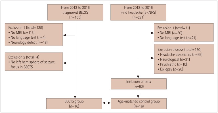
This study included 16 children who were diagnosed with BECTS in the dominant hemisphere and 16 age-matched children as controls. Among 155 patients diagnosed with BECTS, 113 and 4 children were excluded due to MRI with DTI and language testing not being conducted, respectively. A further 18 patients were excluded due to neurological defects that could influence DTI, as were 4 patients with BECTS whose seizure location was not in the dominant hemisphere.
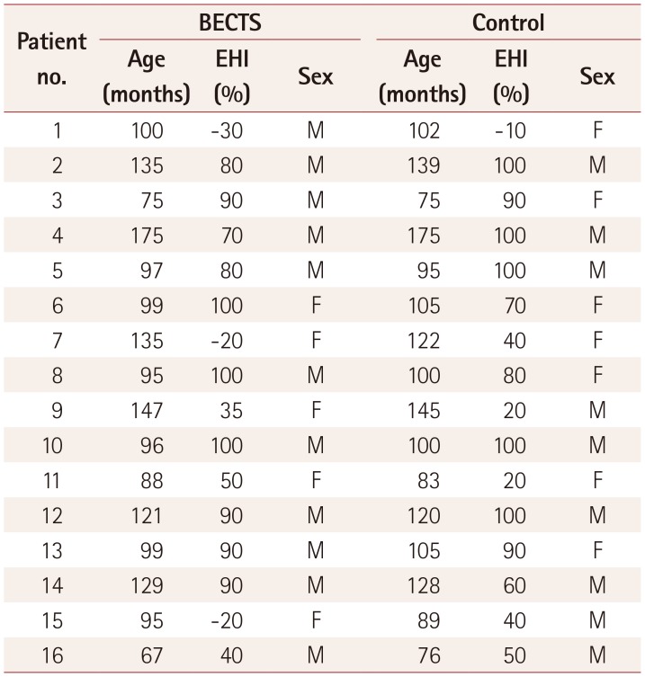
The subjects in the BECTS group were aged 109.6±28.3 months (range, 70–175 months), while those in the control group were aged 108.4±25.9 months (range, 75–175 months). In The EHI revealed right handedness in 10 (62.5%) and ambidexterity in 6 (37.5%) of the subjects in the BECTS and control groups, with no cases of left handedness. There were seven females (31.2%) in the BECTS group and seven (43.8%) in the control group. There were no significant intergroup differences in the demographic characteristics (Fig. 2, Table 1).
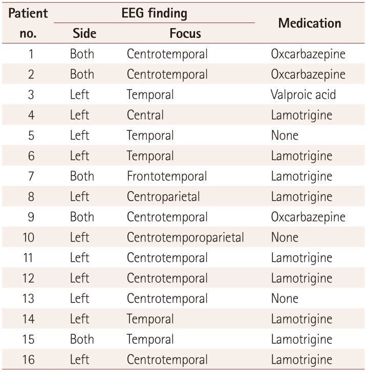
Twelve of the 16 BECTS children had seizure foci in the left hemisphere, and 5 children had seizure foci in both hemispheres. Immediately after the diagnosis, the BECTS patients received antiepileptic drugs if indicated, such as in recurrent cases (Table 2).

Diffusion-tensor magnetic resonance imaging

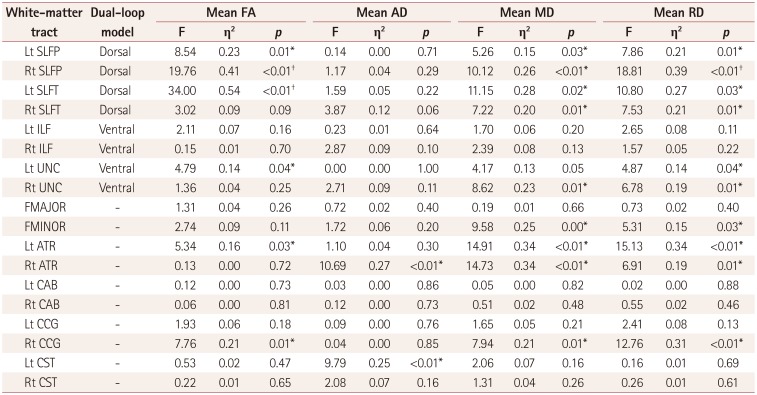
Among the 18 major white-matter tracts extracted by the TRACULA tool in FreeSurfer 5.3 software, the language-related tracts were the SLFP, SLFT, UNC, and ILF. In general, the mean FA corresponds to the inverse proportion of the mean MD and RD, and in this study the BECTS group exhibited a lower mean FA and higher mean MD and RD than controls in the language-related white-matter tracts.
In the dorsal streams, among DTI scalar values of the SLF tracts, the mean FA, MD, and RD differed between the BECTS and control groups. In the dominant hemisphere, which is associated with language, both the SLFP and SLFT showed low mean FA values, with the difference being significant in the BECTS group. In addition, the mean FA for the SLFP tract in the nondominant hemisphere was lower in the BECTS group than in the control group. The ventral streams included the UNC and ILF tracts. The mean FA for the UNC in the dominant hemisphere was significantly lower in the BECTS group than in the control group. In contrast, the mean MD and RD for the UNC were high in the BECTS group. Unlike for the UNC, these values for the ILF did not differ between the two groups. Although not included in the dual-loop model, the left ATR and right CCG also showed a lower mean FA and higher mean RD in the BECTS group than in the control group (Table 3, Fig. 3).

Language tests

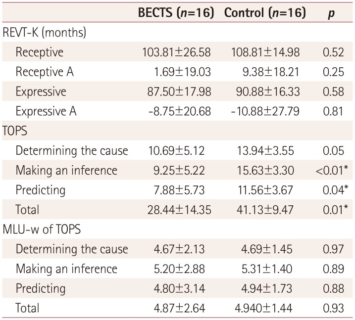
The total score for the TOPS language test was lower in the BECTS group than in the control group (Table 4). Differences were significant for the making-an-inference and predicting subcomponents of the TOPS language test. MLU-w of TOPS and REVT-K was not statistically significant difference.

DISCUSSION

The mean FA of language-related white-matter tracts in the dual-loop model was lower in the BECTS group than in the control group, particularly for the SLF and UNC of the dominant hemisphere. The mean RDs for the SLF and UNC were increased in the BECTS group. The FA of DTI scalar values expresses the degree of this anisotropic state,28 with a value closer to 1 indicating a more-anisotropic state in axon fibers with a specific direction. The present findings therefore support previous descriptions of the language dysfunction of BECTS patients.1
The AF is one of the most important language-related white-matter tracts, and connects the posterior superior temporal gyrus to the inferior frontal gyrus. The SLF is one of the components of the dorsal stream of the dual-loop model, and is located in the white matter connecting the temporoparietal area to the ipsilateral frontal and opercular areas.29 This tract is subdivided into three major temporoparietal subareas: the superior parietal lobe, angular gyrus, and supramarginal gyrus.30 It has recently been suggested that the AF is part of the SLF.31 Among these three subareas, the supramarginal gyrus is related to the language function of articulation.32
FreeSurfer software was used to divide the SLF into the parietal bundle of the SLFP and the temporal bundle of the SLFT. The SLFP and SLFT correspond most closely to the supramarginal gyrus and AF, respectively.33 DTI scalar values were altered in the dominant hemisphere of the SLFT and SLFP tracts in the BECTS group, indicating a relation between BECTS and language dysfunction. Language function mainly occurs in the left brain hemisphere after lateralization. It could be inferred that decreases in the mean FA of language-related white-matter tracts in the dominant hemisphere are associated with lateralization.34
The origin of seizures in BECTS is the Rolandic area located around the central sulcus, namely in the precentral and postcentral gyri of the brain. The SLF and AF are located near the Rolandic area.29 DTI scalar values of FA have been reported to be decreased in the precentral and postcentral gyri, which are the foci of seizure activities in BECTS patients.11 In the present study, the BECTS group exhibited altered DTI scalar values with decreased mean FA and increased RD for the SLFP and SLFT in the dominant hemisphere, which are the areas related to the seizure focus of BECTS patients. These findings provide evidence of a correlation between BECTS and language dysfunction. If seizure activity decreases after appropriate treatment, resulting in the restoration of language ability, the hypothesis could be supported. Hence, further studies of DTI changes after treatment in BECTS patient are needed.
The UNC is a hook-shaped bundle that links the temporal lobe to the inferior frontal gyrus and frontal lobe, and mainly constitutes the ventral stream of the dual-loop model.4 The UNC is also located near the Rolandic area. The findings of a decreased mean FA and increased mean RD for the UNC in the dominant hemisphere in this study could be relevant to its location in the brain. The UNC could be damaged by the spreading seizure activity in BECTS patients. In spite of not being a language-related white-matter tract, the ATR in the dominant hemisphere showed the same result for the DTI scalar value in the language-related white-matter tracts. The ATR connects anterior and midline nuclear groups of the thalamus with the frontal lobe. The ATR pathway included the Rolandic area, which is the seizure focus of BECTS, and so seizure activity in BECTS patients could influence the ATR.
In addition to seizure activity, the pathophysiology of disease could result in the alteration of DTI scalar values in BECTS patients. There are three main causes for FA decreasing in DTI: degradation or congenital abnormalities of the axonal membrane and myelin, and decreased density of myelinated axons.353637 Depending on the age at onset and benign course of BECTS, the cause of BECTS could be related to maturation impairment of the brain.38 Besides seizure activity, the decreased FA value in BECTS is most likely due to the demyelination or decreased density of myelinated axons. Brain impairment in BECTS could decrease the FA value.
In the dual-loop model, the function of the SLF in language was phonological processing.4 The TOPS language test evaluated the logical processing of patients. The decreased FA value and decreased TOPS score for the language test in the BECTS group could imply that BECTS patients have impaired language function in the SLF. A clear correlation between language test results and DTI scalar values could make this inference certain, and so further studies of the relationship between language abilities and DTI scalar values are warranted.
In conclusion, seizure activities are predicted to alter the language ability of BECTS patients. Furthermore, given the lower TOPS scores in the BECTS group, the language defects of BECTS may be more strongly correlated with logical processing.
The main limitations of this study were its retrospective design and the age of the participants meaning that their brains were in a period of rapid development. A previous study of the white-matter tracts using DTI found that the dorsal and ventral streams in the dual-loop model are nearly completely developed in neonates and at 5 years old, respectively.39 The inclusion of school-age subjects could be supplemented by matching the age and the period of development in the dual-loop model. Notwithstanding these limitations, the results of this study are important since they can be utilized when evaluating the language abilities of BECTS patients.

Acknowledgements

The authors thank Prof. Moon-Soo Lee of Korea University for providing the basic concept of DTI, Prof. Chan-Uhng Joo and Prof. Hyo-Jong Lee of Chonbuk National University for their critical reviews of the manuscript, and Mi-Kyung Song (a speech therapist at Chonbuk National University) for helping with the language test.

Notes

Author Contributions:

  • Conceptualization: Sun Jun Kim.

  • Data curation: Sun Jun Kim, Hyun Ho Kim.

  • Investigation: Sun Jun Kim, Hyun Ho Kim.

  • Methodology: Sun Jun Kim, Hyun Ho Kim.

  • Software: Hyun Ho Kim.

  • Supervision: Sun Jun Kim.

  • Writing—original draft: Sun Jun Kim, Hyun Ho Kim.

  • Writing—review & editing: Sun Jun Kim, Gyung Ho Chung, Sung Hee Park.

Conflicts of Interest: The authors have no potential conflicts of interest to disclose.

References

1. Northcott E, Connolly AM, Berroya A, Sabaz M, McIntyre J, Christie J, et al. The neuropsychological and language profile of children with benign rolandic epilepsy. Epilepsia. 2005; 46:924–930. PMID: 15946332.
crossref
2. Völkl-Kernstock S, Bauch-Prater S, Ponocny-Seliger E, Feucht M. Speech and school performance in children with benign partial epilepsy with centro-temporal spikes (BCECTS). Seizure. 2009; 18:320–326. PMID: 19249229.
crossref
3. Filippini M, Ardu E, Stefanelli S, Boni A, Gobbi G, Benso F. Neuropsychological profile in new-onset benign epilepsy with centrotemporal spikes (BECTS): focusing on executive functions. Epilepsy Behav. 2016; 54:71–79. PMID: 26667848.
crossref
4. Rijntjes M, Weiller C, Bormann T, Musso M. The dual loop model: its relation to language and other modalities. Front Evol Neurosci. 2012; 4:9. PMID: 22783188.
crossref
5. Démonet JF, Thierry G, Cardebat D. Renewal of the neurophysiology of language: functional neuroimaging. Physiol Rev. 2005; 85:49–95. PMID: 15618478.
crossref
6. Conturo TE, Lori NF, Cull TS, Akbudak E, Snyder AZ, Shimony JS, et al. Tracking neuronal fiber pathways in the living human brain. Proc Natl Acad Sci U S A. 1999; 96:10422–10427. PMID: 10468624.
crossref
7. Alexander AL, Lee JE, Lazar M, Boudos R, DuBray MB, Oakes TR, et al. Diffusion tensor imaging of the corpus callosum in Autism. Neuroimage. 2007; 34:61–73. PMID: 17023185.
crossref
8. Ameis SH, Lerch JP, Taylor MJ, Lee W, Viviano JD, Pipitone J, et al. A diffusion tensor imaging study in children with ADHD, autism spectrum disorder, OCD, and matched controls: distinct and non-distinct white matter disruption and dimensional brain-behavior relationships. Am J Psychiatry. 2016; 173:1213–1222. PMID: 27363509.
crossref
9. Friederici AD. Pathways to language: fiber tracts in the human brain. Trends Cogn Sci. 2009; 13:175–181. PMID: 19223226.
crossref
10. Sundgren PC, Dong Q, Gómez-Hassan D, Mukherji SK, Maly P, Welsh R. Diffusion tensor imaging of the brain: review of clinical applications. Neuroradiology. 2004; 46:339–350. PMID: 15103435.
crossref
11. Ciumas C, Saignavongs M, Ilski F, Herbillon V, Laurent A, Lothe A, et al. White matter development in children with benign childhood epilepsy with centro-temporal spikes. Brain. 2014; 137:1095–1106. PMID: 24598359.
crossref
12. Good CD, Johnsrude IS, Ashburner J, Henson RN, Friston KJ, Frackowiak RS. A voxel-based morphometric study of ageing in 465 normal adult human brains. Neuroimage. 2001; 14:21–36. PMID: 11525331.
crossref
13. Afzali M, Soltanian-Zadeh H, Elisevich KV. Tract based spatial statistical analysis and voxel based morphometry of diffusion indices in temporal lobe epilepsy. Comput Biol Med. 2011; 41:1082–1091. PMID: 21616484.
crossref
14. Xiao F, Chen Q, Yu X, Tang Y, Luo C, Fang J, et al. Hemispheric lateralization of microstructural white matter abnormalities in children with active benign childhood epilepsy with centrotemporal spikes (BECTS): a preliminary DTI study. J Neurol Sci. 2014; 336:171–179. PMID: 24210075.
crossref
15. Cao W, Zhang Y, Hou C, Yang F, Gong J, Jiang S, et al. Abnormal asymmetry in benign epilepsy with unilateral and bilateral centrotemporal spikes: a combined fMRI and DTI study. Epilepsy Res. 2017; 135:56–63. PMID: 28623837.
crossref
16. Liamsuwan S, Grattan-Smith P, Fagan E, Bleasel A, Antony J. The value of partial sleep deprivation as a routine measure in pediatric electroencephalography. J Child Neurol. 2000; 15:26–29. PMID: 10641606.
crossref
17. Oldfield RC. The assessment and analysis of handedness: the Edinburgh inventory. Neuropsychologia. 1971; 9:97–113. PMID: 5146491.
crossref
18. Reuter M, Rosas HD, Fischl B. Highly accurate inverse consistent registration: a robust approach. Neuroimage. 2010; 53:1181–1196. PMID: 20637289.
crossref
19. Fischl B, Salat DH, Busa E, Albert M, Dieterich M, Haselgrove C, et al. Whole brain segmentation: automated labeling of neuroanatomical structures in the human brain. Neuron. 2002; 33:341–355. PMID: 11832223.
20. Fischl B, Dale AM. Measuring the thickness of the human cerebral cortex from magnetic resonance images. Proc Natl Acad Sci U S A. 2000; 97:11050–11055. PMID: 10984517.
crossref
21. Sled JG, Zijdenbos AP, Evans AC. A nonparametric method for automatic correction of intensity nonuniformity in MRI data. IEEE Trans Med Imaging. 1998; 17:87–97. PMID: 9617910.
crossref
22. Yendiki A, Panneck P, Srinivasan P, Stevens A, Zöllei L, Augustinack J, et al. Automated probabilistic reconstruction of white-matter pathways in health and disease using an atlas of the underlying anatomy. Front Neuroinform. 2011; 5:23. PMID: 22016733.
crossref
23. Andersson JLR, Sotiropoulos SN. An integrated approach to correction for off-resonance effects and subject movement in diffusion MR imaging. Neuroimage. 2016; 125:1063–1078. PMID: 26481672.
crossref
24. Yendiki A, Koldewyn K, Kakunoori S, Kanwisher N, Fischl B. Spurious group differences due to head motion in a diffusion MRI study. Neuroimage. 2014; 88:79–90. PMID: 24269273.
crossref
25. Kim YT, Hong GH, Kim KH. Content and reliability analyses of the receptive and expressive vocabulary test (REVT). Commun Sci Disord. 2009; 14:34–45.
26. Bae SY, Lim SS, Lee JH. Test of problem solving. Seoul: Seoul Community Rehabilitation Center;2000.
27. Lee HJ, Kim YT. Measures of utterance length of normal language-delayed children. Commun Sci Disord. 1999; 4:1–14.
28. Pfefferbaum A, Sullivan EV, Hedehus M, Lim KO, Adalsteinsson E, Moseley M. Age-related decline in brain white matter anisotropy measured with spatially corrected echo-planar diffusion tensor imaging. Magn Reson Med. 2000; 44:259–268. PMID: 10918325.
29. Madhavan KM, McQueeny T, Howe SR, Shear P, Szaflarski J. Superior longitudinal fasciculus and language functioning in healthy aging. Brain Res. 2014; 1562:11–22. PMID: 24680744.
crossref
30. Schmahmann JD, Pandya DN, Wang R, Dai G, D'Arceuil HE, De Crespigny AJ, et al. Association fibre pathways of the brain: parallel observations from diffusion spectrum imaging and autoradiography. Brain. 2007; 130:630–653. PMID: 17293361.
crossref
31. Yeterian EH, Pandya DN, Tomaiuolo F, Petrides M. The cortical connectivity of the prefrontal cortex in the monkey brain. Cortex. 2012; 48:58–81. PMID: 21481342.
crossref
32. Makris N, Kennedy DN, McInerney S, Sorensen AG, Wang R, Caviness VS Jr, et al. Segmentation of subcomponents within the superior longitudinal fascicle in humans: a quantitative, in vivo, DT-MRI study. Cereb Cortex. 2005; 15:854–869. PMID: 15590909.
crossref
33. Catani M, Thiebaut de. A diffusion tensor imaging tractography atlas for virtual in vivo dissections. Cortex. 2008; 44:1105–1132. PMID: 18619589.
crossref
34. Dubois J, Hertz-Pannier L, Cachia A, Mangin JF, Le Bihan D, Dehaene-Lambertz G. Structural asymmetries in the infant language and sensori-motor networks. Cereb Cortex. 2009; 19:414–423. PMID: 18562332.
crossref
35. Pierpaoli C, Barnett A, Pajevic S, Chen R, Penix LR, Virta A, et al. Water diffusion changes in Wallerian degeneration and their dependence on white matter architecture. Neuroimage. 2001; 13:1174–1185. PMID: 11352623.
crossref
36. Song SK, Sun SW, Ramsbottom MJ, Chang C, Russell J, Cross AH. Dysmyelination revealed through MRI as increased radial (but unchanged axial) diffusion of water. Neuroimage. 2002; 17:1429–1436. PMID: 12414282.
crossref
37. Takahashi M, Hackney DB, Zhang G, Wehrli SL, Wright AC, O'Brien WT, et al. Magnetic resonance microimaging of intraaxonal water diffusion in live excised lamprey spinal cord. Proc Natl Acad Sci U S A. 2002; 99:16192–16196. PMID: 12451179.
crossref
38. Chahine LM, Mikati MA. Benign pediatric localization-related epilepsies. Epileptic Disord. 2006; 8:243–258. PMID: 17150437.
39. Brauer J, Anwander A, Perani D, Friederici AD. Dorsal and ventral pathways in language development. Brain Lang. 2013; 127:289–295. PMID: 23643035.
crossref
Fig. 1

Imaging view of the 18 reconstructed cerebral tracts in a control subject. A: Coronal view. B: Sagittal view. C: Axial view. D: Three-dimensional view. ATR: anterior thalamic radiation, CAB: cingulum-angular bundle, CCG: cingulum-cingulate-gyrus bundle, CST: cortical spinal tract, FMAJOR: corpus callosum-forceps major, FMINOR: corpus callosum-forceps minor, ILF: inferior longitudinal fasciculus, SLFP: superior longitudinal fasciculus-parietal terminations, SLFT: superior longitudinal fasciculus-temporal terminations, UNC: uncinate fasciculus.

jcn-15-502-g001
Fig. 2

Flow chart of the study participants. BECTS: benign epilepsy with centrotemporal spikes, NRS: numeric rating scale.

jcn-15-502-g002
Fig. 3

Comparison of diffusion-tensor MRI scalar values of the 18 major white-matter tracts in the BECTS and control groups. Data indicate group mean and standard-error values. A: Mean FA of BECTS group was decreased in dorsal stream of language related white-matter-tract. B: There was no difference between control and BECTS groups in mean AD. C and D: Mean MD and RD of BECTS group were increased in dorsal stream of language related white-matter-tract. *Significant at p<0.05 (uncorrected), Significant correlation after Bonferroni correction (p<0.05/18=0.003). AD: axial diffusivity, ATR: anterior thalamic radiation, BECTS: benign epilepsy with centrotemporal spikes, CAB: cingulum-angular bundle, CCG: cingulum-cingulate gyrus bundle, CST: cortical spinal tract, FA: fractional anisotropy, FMAJOR: corpus callosum-forceps major, FMINOR: corpus callosum-forceps minor, ILF: inferior longitudinal fasciculus, Lt: left, MD: mean diffusivity, RD: radial diffusivity, Rt: right, SLFP: superior longitudinal fasciculus-parietal terminations, SLFT: superior longitudinal fasciculus-temporal terminations, UNC: uncinate fasciculus.

jcn-15-502-g003
Table 1

Demographic characteristics in the two study groups

jcn-15-502-i001

BECTS: benign epilepsy with centrotemporal spikes, EHI: Edinburgh Handedness Inventory, F: female, M: male.

Table 2

EEG findings and medications in the benign epilepsy with centrotemporal spikes group

jcn-15-502-i002
Table 3

Comparison of diffusion-tensor MRI scalar values between the BECTS and control groups

jcn-15-502-i003

*Significant at p<0.05 (uncorrected), Significant correlation after Bonferroni correction (p<0.05/18=0.003).

η2: effect size, AD: axial diffusivity, ATR: anterior thalamic radiation, BECTS: benign epilepsy with centrotemporal spikes, CAB: cingulum-angular bundle, CCG: cingulum-cingulate gyrus bundle, CST: cortical spinal tract, FA: fractional anisotropy, FMAJOR: corpus callosum-forceps major, FMINOR: corpus callosum-forceps minor, ILF: inferior longitudinal fasciculus, Lt: left, MD: mean diffusivity, RD: radial diffusivity, Rt: right, SLFP: superior longitudinal fasciculus-parietal termination, SLFT: superior longitudinal fasciculus-temporal termination, UNC: uncinate fasciculus.

Table 4

Comparison of language test results between the BECTS and control groups

jcn-15-502-i004

*Significant at p<0.05.

BECTS: benign epilepsy with centrotemporal spikes, Expressive A: chronological age-expressive age, MLU-w: mean length of utterance in words, Receptive A: chronological age-receptive age, REVT-K: Korean version of the Receptive and Expressive Vocabulary Test, TOPS: Test of Problem-Solving Abilities.

TOOLS
Similar articles