

 PDF
PDF ePub
ePub Citation
 Print
Print


Ji-Eun Kim1 , In-Gyeong Yun1, Seong-Soog Jeong1, 2, Ki-Ho Chung1, 2, Choong-Ho Choi1, 2
, In-Gyeong Yun1, Seong-Soog Jeong1, 2, Ki-Ho Chung1, 2, Choong-Ho Choi1, 2
Abstract
Objectives
We examined the effect of commercial plum beverages on dental erosion and whether the addition of calcium to these beverages would inhibit dental erosion.
Methods
We analyzed three groups as follows: Maesil 1 group (Chorok Maesil), Maesil 2 group (Sunkist plum), both of which were selected from commercially-available plum beverages, and Calcium-added maesil group (addition of 3% calcium to Chorok Maesil). For negative and positive control groups, Jeju Samdasoo and Coca Cola were selected, respectively. The characteristics of the experimental beverages were analyzed, and the specimens were immersed in the experimental beverage. The degree of erosion was measured by Vickers hardness number (VHN) and scanning electron microscope images.
Results
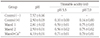
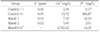
Positive control group had the lowest pH (2.50±0.03), followed by Maesil 2 (pH 2.59±0.01), Maesil 1 (pH 2.81±0.02), calcium-added maesil (pH 4.19±0.01), and negative control group (pH 7.57±0.06). Significant differences were found in surface microhardness between positive control, Maesil 1, Maesil 2 and calcium-added maesil group before immersion and at 30 minutes after immersion (P<0.05), and change in VHN (positive control group, −80.94±20.63; Maesil 1 group, −69.33±24.88; and Maesil 2 group, −78.49±18.60 in comparison with negative control group, −6.57±26.73). There was no significant difference (P<0.05) in change in VHN between calcium-added maesil (−13.02±17.33) and negative control group.
Conclusions
Plum beverages can potentially induce dental erosion due to their low pH. However, adding calcium to these beverages can reduce the risk of dental erosion. Therefore, the risk of dental erosion must be considered during consumption of plum beverages, and the addition of calcium into plum beverages may be considered as a way to prevent dental erosion.
Figures and Tables
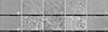 | Fig. 1SEM images of enamel surface after beverage treatment for 30 minutes (1, Control (−); 2, Control (+); 3, Maesil 1; 4, Maesil 2; 5, Maesil+Ca2+; a, ×10,000; b, ×50,000).
|


References
1. Shin HL. Consumer attitude survey : Beverage purchasing behaviors and preferences [master's thesis]. Seoul: Sejong University;2010. [Korean].
2. Jun MK, Lee DH, Lee SM. Assessment of nutrient and sugar content and pH of some commercial beverages. J Dent Hyg Sci. 2016; 16:464–471.

3. The Food&beverage News. [Internet]. The Food&beverage News. cited 2019 Aug 28. Available from: https://www.thinkfood.co.kr/news/articleView.html?idxno=6977.
4. Shim JH, Park MW, Kim MR, Lim KT, Park ST. Screening of antioxidants in fructus mume (Prunus mume Sieb. et Zucc.) extract. Agric Chem Biotechnol. 2002; 45:119–123.
5. Lee JS, Chung KH. Antimicrobial effect of Prunus mume extracts against cariogenic bacteria. J Korean Acad Oral Health. 2017; 41:65–70.
6. Jang JH, Kim YI, Lee H. Antimicrobial activity of Prunus mume extract to oral microbes. J Korean Soc Dent Hyg. 2014; 14:109–115.
7. Lee DS, Woo SK, Yang CB. Studies on chemical composition of major fruits in Korea : On non-volatile organic acid and sugar contents of apricot (maesil), peach, grape, apple and pear and its seasonal variation. Korean J Food Sci Technol. 1972; 4:134–139.
8. ten Cate JM, Imfeld T. Dental erosion, summary. Eur J Oral Sci. 1996; 104(2 Pt 2):241–244.
9. Zero DT. Etiology of dental erosion—extrinsic factors. Eur J Oral Sci. 1996; 104(2 Pt 2):162–177.
10. Lussi A, Schaffner M. Progression of and risk factors for dental erosion and wedge-shaped defects over a 6-year period. Caries Res. 2000; 34:182–187.
11. Choi DY, Shin SC. A study on pH of several beverages in Korea. J Korean Acad Dent Health. 1996; 20:399–410.
12. Lee HJ. Erosive effect of hangover beverages on bovine teeth [Master's thesis]. Gwangju: Chonnam National University;2011. [Korean].
13. Chunmuang S, Jitpukdeebodintra S, Chuenarrom C, Benjakul P. Effect of xylitol and fluoride on enamel erosion in vitro. J Oral Sci. 2007; 49:293–297.
14. Ramalingam L, Messer LB, Reynolds EC. Adding casein phosphopeptide-amorphous calcium phosphate to sports drinks to eliminate in vitro erosion. Pediatr Dent. 2005; 27:61–67.
15. West NX, Hughes JA, Parker DM, Newcombe RG, Addy M. Development and evaluation of a low erosive blackcurrant juice drink 2. Comparison with a conventional blackcurrant juice drink and orange juice. J Dent. 1999; 27:341–344.
16. Scheutzel P. Etiology of dental erosion-intrinsic factors. Eur J Oral Sci. 1996; 104(2 Pt 2):178–190.
17. Johansson AK, Lingström P, Birkhed D. Comparison of factors potentially related to the occurrence of dental erosion in high-and low-erosion groups. Eur J Oral Sci. 2002; 110:204–211.
18. Attin T, Meyer K, Hellwig E, Buchallar W, Lennon AM. Effect of mineral supplements to citric acid on enamel erosion. Arch Oral Biol. 2003; 48:753–759.
19. Larsen MJ. Prevention by means of fluoride of enamel erosion as caused by soft drinks and orange juice. Caries Res. 2001; 35:229–234.
20. Larsen MJ, Nyvad B. Enamel erosion by some soft drinks and orange juices relative to their pH, buffering effect and contents of calcium phosphate. Caries Res. 1999; 33:81–87.
21. Shellis RP, Barbour ME, Jesani A, Lussi A. Effects of buffering properties and undissociated acid concentration on dissolution of dental enamel in relation to pH and acid type. Caries Res. 2013; 47:601–611.
22. Thylstrup A, Fejerskov O. Textbook of clinical cariology. 2nd ed. Copenhagen: Munksgaard;1994. p. 231–257. p. 288–299.
23. Rytömaa I, Meurman JH, Koskinen J, Laakso T, Gharazi L, Turunen R. In vitro erosion of bovine enamel caused by acidic drinks and other foodstuffs. Scand J Dent Res. 1988; 96:324–333.
24. Ji SG. New Food Additive theory and reality. 3th ed. Seoul: The Food Journal;2008. p. 114–115.
25. Lee HJ, Oh HN, Hong SJ, Choi CH. Effect of hangover beverage containing fluoride and calcium on enamel erosion. J Korean Acad Oral Health. 2012; 36:177–184.
26. Gregory-Head B, Curtis DA. Erosion caused by gastroesophageal reflux: diagnostic considerations. J Prosthodont. 1997; 6:278–285.
27. Ahn HY, Lee KH, Kim DE. Erosion of tooth enamel by acidic drinks and remineralization by artificial saliva. J Korean Acad Pediatr Dent. 2002; 29:84–91.
28. Woltgens JH, Vingerling P, de Blieck-Hogervorst JM, Bervoets DJ. Enamel erosion and saliva. Clin Prev Dent. 1985; 7:8–10.
29. Amaechi BT, Higham SM, Edgar WM, Milosevic A. Thickness of acquired salivary pellicle as a determinant of the sites of dental erosion. J Dent Res. 1999; 78:1821–1828.
30. Shim JH, Jeong TS, Kim S. A study on the enamel erosion by fermented milks. J Korean Acad Pediatr Dent. 2004; 31:555–563.
 XML Download