

 PDF
PDF ePub
ePub Citation
 Print
Print


Introduction
Varicella Zoster Virus (VZV) infection is usually a self-limited disease in immunocompetent patients; however, it could be fatal for immunocompromised patients; such as organ transplantation recipients [1]. Theoretically, intravenous immunoglobulin (IVIG) could suppress viremia by preventing invasion of VZV into target cells [23]. However, there are few reports on whether IVIG is beneficial in immunocompromised patients with severe varicella [34]. Here, we describe a case of a young adult kidney transplant recipient who had severe varicella with fulminant hepatitis and was successfully treated by intravenous acyclovir (ACV) with adjuvant IVIG. We present the details of viral kinetics in the plasma and of humoral and cell-mediated immune responses from this case.
Case report
A 21-year-old woman who had received a living-related kidney transplant 1 year earlier presented with fever and generalized rash for 5 days. She had a generalized multistage blister-like rash (Supplemental Fig. 1). She was admitted to another hospital and was diagnosed with chickenpox. She received ACV 10 mg/kg IV every 12 hours for 2 days according to her estimated glomerular filtration rate of 38 ml/min/1.73m2. However, the fulminant hepatitis progressed and she was referred to our hospital for an emergency liver transplantation. Her immunosuppressive regimen consisted of tacrolimus 2 mg twice a day, mycophenolate mofetil 1 g twice a day, and prednisone 7.5 mg once daily. Her antimicrobial prophylaxis consisted of trimethoprim/sulfamethoxazole. She had no past history of chickenpox, and had received varicella vaccine at 12 months of age. She did not drink alcohol, and there was no history of taking herbal medicine. Initial blood examination revealed a white blood cell count of 22.8×10³/mm3, hemoglobin 13.3 g/dL, platelet count 53×103/mm3, prothrombin time (PT) 17.8 sec, serum creatinine 1.65 mg/dL, alanine aminotransferase (ALT) 2,016 IU/L, aspartate aminotransferase (AST) 4,434 IU/L, albumin 4.1 g/dL, blood urea nitrogen (BUN) 20 mg/dL, total bilirubin 0.4 mg/dL, lactate dehydrogenase (LD) 6,484 IU/L, and creatine kinase (CK) 117 IU/L. Serum examination for hepatitis B virus (HBV), hepatitis C virus (HCV), human immunodeficiency virus (HIV), and cytomegalovirus(CMV) were negative, and additional workup including autoimmune and Wilson's disease was unremarkable. Initial IgM and IgG for VZV were negative on hospital day (HD 1), but VZV-specific PCR from skin vesicles and blood was positive on HD 7. Thus, the patient was diagnosed with varicella with fulminant hepatitis. The patient received empirical antibiotics including meropenem 1 g IV every 12 hours from HD 1 to HD 6, teicoplanin 400 mg IV every 72 hours from HD 1 to HD 3, ACV 10 mg/kg IV every 12 hours according to her estimated glomerular filtration rate of 38 ml/min/1.73m2 from HD 1 to HD 15. Because the initial trough levels of tacrolimus was 13.7 ng/mL, the tacrolimus dose was decreased to 1 mg twice a day with a target trough concentration range of 6-8 ng/mL; prednisone dose was decreased to 5 mg once daily, and mycophenolate mofetil was discontinued. On HD 2, her ALT and AST rapidly increased to 7,638 IU/L and 3,034 IU/L, respectively, and PT increased to 25.8 sec. Adjuvant IVIG 500 mg/kg/day was administered from HD 2 to 7. The decreasing slope (beta coefficient −0.446) of blood viral load was steeper during the IVIG therapy than after the therapy(beta coefficient −0.123) (P = 0.04, Fig. 1A), and VZV glycoprotein IgG titer and VZV-specific T cell response were not detectible during the 5-day IVIG therapy (Fig. 1B and 1C). On HD 7, her AST and ALT decreased to 265 IU/L and 435 IU/L, respectively, and PT decreased to 16.2 sec; however, platelet count decreased to 6x10³/mm3 and serum creatinine increased to 2.49 mg/dL. Her general condition and laboratory findings improved and she was discharged without any complication on HD 14.
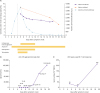 | Figure 1(A) Detailed kinetic data on varicella-zoster virus (VZV) DNA loads in plasma and saliva, and alanine aminotransferase, according to days after symptom onset in a 21-year-old woman with fulminant varicella hepatitis. The decline in viral copies in plasma (pupple line) and saliva (orange line), and alanine aminotransferase (sky blue line) are shown. The duration of antimicrobial therapy and intravenous immunoglobulin(IVIG) is shown by the yellow bars. The dashed line represents the day of hospitalization. (B) Changes of VZV glycoprotein IgG titer and (C) VZV lysate-specific T cell response as a function of hospital day. (PBMCs: peripheral blood mononuclear cells).
|
1. Varicella zoster viral load
To quantify the viral load, patient's plasma and saliva were collected. Saliva samples of one mL or more were collected using Omnigene-Oral kit (DNA GenotekInc., Ottawa, Ontario, Canada) at any time of day, but at least one hour after a meal. The saliva samples were vigorously shaken for at least 10 seconds and incubated in a water bath at 50 °C for 1 hour. DNA was extracted from plasma and saliva samples using a Qia-Amp DNA mini kit (Qiagen Inc., Chatsworth, CA, USA), and VZV was quantified with a VZV real-time PCR kit (GeneProof, Brno, Czech Republic) using a LightCycler 480 System (Roche, Basel, Switzerland). VZV DNA copy number was determined by comparing the cycle threshold (Ct) value of test samples with the Ct value of the reference VZV DNA provided with the PCR kit. The limit of quantitative PCR detection was 1 copy of VZV DNA per PCR reaction or 10 copies per mL of plasma or saliva.
2. VZV-specific T cell response and antibody response
Peripheral blood mononuclear cells (PBMCs) were isolated from whole blood samples using lymphocyte separation medium (Corning Inc., New York, NY, USA) and cryopreserved. Cryopreserved PBMCs were quickly thawed at 37°C, washed twice with RPMI-1640 medium with 10% heat-inactivated fetal bovine serum, and resuspended at 2 × 10⁶ cells/mL. Samples of 100 μL of PBMCs were seeded onto 96-well plates coated with anti-human interferon gamma antibody (Thermo Fisher Scientific, Waltham, MA, USA, 2 µg/mL) and stimulated with a VZV lysate (VZ-10 strain, Microbix Biosystems Inc. Toronto, Ontario, Canada). Phytohemagglutinin (Sigma Aldrich, St. Louis, MO, USA; 50 ng/mL) was used as a positive control. Spots were counted and analyzed with an automated enzyme-linked ImmunoSpot plate reader (ImmunoSpot analyzer, Cellular Technology Limited, Shaker Heights, Cleveland, OH, USA). The resulting spot numbers were expressed as mean numbers of spot-forming units per 106 cells in duplicate assays. The titers of anti-VZV glycoprotein IgG in plasma samples from the patient were measured with a commercial VZV glycoprotein ELISA kit (Binding Site Group Ltd, Edgbaston, Birmingham, UK).
3. Statistical Analysis
The patient's plasma viral loads (varicella copies/mL) were plotted on a logarithmic scale against time (in days) and fitted by linear regression. We evaluated the slope changes between before and after IVIG administration, which was assessed by adding the interaction effect to linear regression. Statistical significance was defined at P <0.05.
Discussion
Early ACV therapy has been known to prevent dissemination of VZV and ameliorate the disease course [56]. However, in some disseminated VZV infection, ACV may not be sufficient for controlling VZV infection in immunocompromised patients [78]. In the present case, fulminant hepatitis due to VZV progressed despite 2-day ACV therapy, at which point the patient was referred to our hospital. We administered adjuvant IVIG with intravenous ACV therapy, and her condition gradually improved. After infusion of IVIG for 5 days, serum creatinine increased. Considering the cause of elevated serum creatinine, IVIG might not be excluded. The use of IVIG must be carried out with care because renal toxicity following IVIG infusion is not uncommon. Also, infusion of IVIG may lead to several complications such as anaphylaxis, central nerve system complications, thromboembolism, hemolysis, and neutropenia [9]. A previous study showed that current IVIG preparations have high levels of VZV-specific IgG despite waning immunity to VZV in the general population due to the lack of circulating virus [10]. However, there are few data on whether IVIG can suppress viremia by neutralizing VZV. Despite continuous ACV infusion, the slope of blood viral load was steeper during the 5-day IVIG infusion than that after the infusion. Furthermore, we clearly showed that VZV-specific T cell and antibody responses were not mounted up to the end of the IVIG infusion (Fig. 1B and 1C), which means that the patient's own immune response was not yet able to control the disseminated VZV during the IVIG therapy. Previous studies have shown that VZV-specific IgG antibody is detected in most patients within the first 4 days after the onset of rash [11]. In addition, viral load in peripheral blood in patients with varicella rapidly decline 1 week after the onset of symptom [12]. We assume that the immunocompromised status may have contributed to the delayed antibody response and viral clearance in this patient. We believe that this case provides important experimental evidence that adjuvant IVIG can significantly reduce viral load kinetics in immunocompromised patients with severe varicella.
 XML Download