

 PDF
PDF ePub
ePub Citation
 Print
Print


INTRODUCTION
Hydatid disease in humans is a parasitic infestation usually caused by the larvae of Echinococcus granulosus (E. granulosus) (1). Although dogs and sheep are the typical hosts of E. granulosus, humans can be opportunistically infected via contaminated water or food containing the parasitic eggs (2). The ingested eggs may penetrate the small bowel wall and then enter the liver through the portal vein (2). Thus, hydatid disease in humans primarily involves the liver (123). This condition is usually asymptomatic and presents with cystic lesions in the infected organs. Among the various complications of hydatid disease, direct rupture of the parasitic cyst into the stomach is extremely rare (2). In this report, we describe a case of a patient with a gastric ulcer that was caused by a ruptured hepatic hydatid cyst.
CASE REPORT
A 49-year-old man visited our hospital with a complaint of upper abdominal pain that had developed a week ago. He had no history of any previous abdominal surgery or trauma. He was a non-drinker and had no history of hepatitis, hypertension, tuberculosis, or diabetes mellitus. He traveled in Uzbekistan a few years ago. On physical examination, the abdomen was found to be soft and flat with no rigidity. However, he complained of discomfort on palpation in the epigastric area. The initial routine laboratory tests revealed the following: hemoglobin level, 10.8 g/dL (normal range, 12–18); white blood cell count, 17.4 × 10³/mm3 (range, 4.8–10.8); and C-reactive protein level, 14.9 mg/dL (range, 0–0.6). The serum levels of aspartate aminotransferase, alanine aminotransferase, alkaline phosphatase, and total bilirubin were within the normal limits. Levels of tumor markers (alpha-fetoprotein, carcinoembryonic antigen, and carbohydrate antigen 19–9) were also within the normal limits.
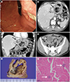
Endoscopic imaging demonstrated a deep ulcer in the lesser curvature side of the gastric upper body surrounded by edematous mucosa (Fig. 1A). An axial CT image showed a round mass, with mottled air bubbles between the hepatic left lateral segment and stomach, causing gastric ulceration (Fig. 1B). On coronal CT images, the mass was located in the hepatic left lateral segment and gastrohepatic ligament, and showed a daughter cyst and peripheral coarse calcifications (Fig. 1C). Further, an additional cystic mass was found in the pelvic cavity (Fig. 1D).
A provisional diagnosis of hydatid disease was made, for which the patient underwent hepatic left lateral sectionectomy, gastric wedge resection, and pelvic mass excision. The gross surgical specimen showed a cystic mass containing pus-like yellow material and calcifications in the hepatic left lateral segment (Fig. 1E), which communicated with the gastric wall through the gastrohepatic ligament. Microscopic examination revealed protoscolices within necrotic debris (Fig. 1F). This histological finding led to the diagnosis of hydatid disease in the liver with spontaneous rupture into the stomach and peritoneal seeding.
DISCUSSION
In humans with hydatid disease, the liver is reported to be the most frequently affected site (approximately 75% of the cases) (2). However, hydatid disease in humans can involve almost any anatomic site, including the pancreas, spleen, kidney, peritoneal cavity, thorax, head, and neck (123). Hydatid disease is usually included in the differential diagnoses in patients with hepatic cystic lesions with calcifications, especially in endemic areas such as the Mediterranean region, the Middle East, Central Asia, sub-Saharan Africa, and South America (3). The parasitic cyst consists of the outer pericyst, middle laminated membrane, and inner germinal layer (1234). Among these three components, the germinal layer plays a central role in the development of the scolices (the larval stage of E. granulosus) and protoscolices (juvenile scolices) (3).
The imaging findings of hydatid disease vary according to its stage of development, ranging from cystic lesions to a completely solid mass (3). The cyst may be unilocular or contain daughter cysts at the periphery of the mother cyst. Hydatid cysts are more frequently accompanied by calcifications in cases involving the liver, spleen, or kidney (1). When present, calcifications are usually distributed along the cyst wall, although internal calcification may also be detected in the hydatid matrix (2). Often, “floating membranes” are observed within the cyst that may appear as serpentine linear structures, a finding that is highly suggestive of hydatid disease (2). In our case, although floating membranes were not typically found within the cyst, the detection of a daughter cyst and coarse calcifications of the cyst wall might be helpful in the correct diagnosis of hydatid disease.
Hepatic hydatid cysts may show exophytic growth through the bare area of the liver and the gastrohepatic ligament (2). Among the many potential local complications of hepatic hydatid cysts, including rupture, infection, biliary communication, and peritoneal seeding, spontaneous rupture of the cyst into the hollow viscera is extremely rare, which may occur in less than 0.5% of the cases (25). The CT images may demonstrate a cyst with air bubbles or an air-fluid level, as observed in our case (6).
Most cases of peritoneal hydatid cyst occur secondary to a hepatic rupture (2). The majority of patients have a history of previous surgery for hepatic hydatid disease, although spontaneous and asymptomatic perforation of hepatic cysts into the peritoneal cavity may also occur (2). Peritoneal hydatid disease may be widespread and involve any site in the abdominopelvic cavity. Its imaging features are analogous to those of hydatid disease involving the liver or spleen. Patients may remain asymptomatic until the cysts are large enough to produce a mass effect on the adjacent viscera (1). With respect to the treatment, surgical resection is preferred and should be followed by adjuvant anthelmintic therapy (1).
In conclusion, we present a case of a patient with hepatic hydatid disease, which caused a gastric ulcer that developed due to spontaneous and direct rupture of the hydatid cyst into the gastric lumen.
 XML Download