

 PDF
PDF ePub
ePub Citation
 Print
Print


Abstract
Cerebral air embolism is a rare, potentially catastrophic iatrogenic complication of central venous catheter removal. Cerebral air embolism can lead to serious neurological sequelae, resulting from cerebral infarction. Early radiological diagnosis of cerebral air embolism is critical for emergent hyperbaric oxygen treatment. In this study, we report the case of a 68-year-old man who developed cerebral air embolism after the removal of a central venous catheter that was immediately diagnosed using brain CT and brain diffusion-weighted imaging.
References
1. Brockmeyer J, Simon T, Seery J, Johnson E, Armstrong P. Cerebral air embolism following removal of central venous catheter. Mil Med. 2009; 174:878–881.

2. Zong Y. Cerebral air embolism after central venous catheter removal: a case report and literature review.J Anesth Crit Care Open Access. 2014; 1:00006.
3. Eum DH, Lee SH, Kim HW, Jung MJ, Lee JG. Cerebral air embolism following the removal of a central venous catheter in the absence of intracardiac right-to-left shunting: a case report.Medicine (Baltimore). 2015; 94:e630.
4. Malhotra K, Rayi A. Gyriform infarction in cerebral air embolism: imaging mimicker of status epilepticus. Ann Indian Acad Neurol. 2017; 20:313–315.
5. Jung HS, Jeong HW, In HS. Cerebral air embolism in a patient with a tuberculous-destroyed lung during commercial air travel: a case report.J Korean Soc Radiol. 2011; 65:109–112.
6. Jeon SB, Kim JS, Lee DK, Kang DW, Kwon SU. Clinicoradiological characteristics of cerebral air embolism. Cerebrovasc Dis. 2007; 23:459–462.
7. Blanc P, Boussuges A, Henriette K, Sainty JM, Deleflie M. Iatrogenic cerebral air embolism: importance of an early hyperbaric oxygenation. Intensive Care Med. 2002; 28:559–563.
Fig. 1.
A 68-year-old man who developed cerebral air embolism after the removal of a central venous catheter. A. Initial non-contrast-enhanced brain CT shows multifocal curvilinear areas of very low attenuation (arrows; about −120 HU) along the right frontal sulcus, suggesting air emboli.
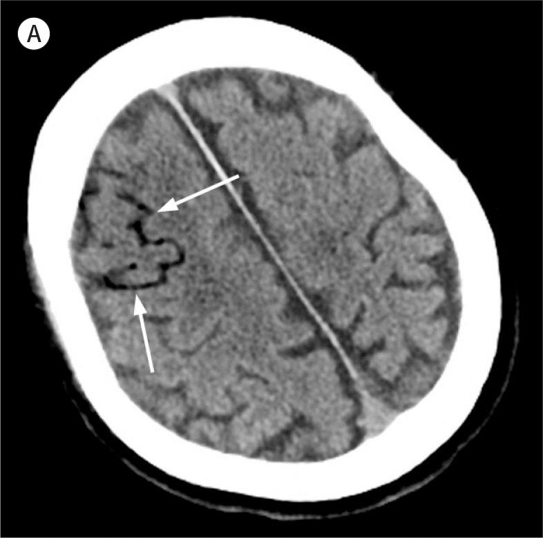
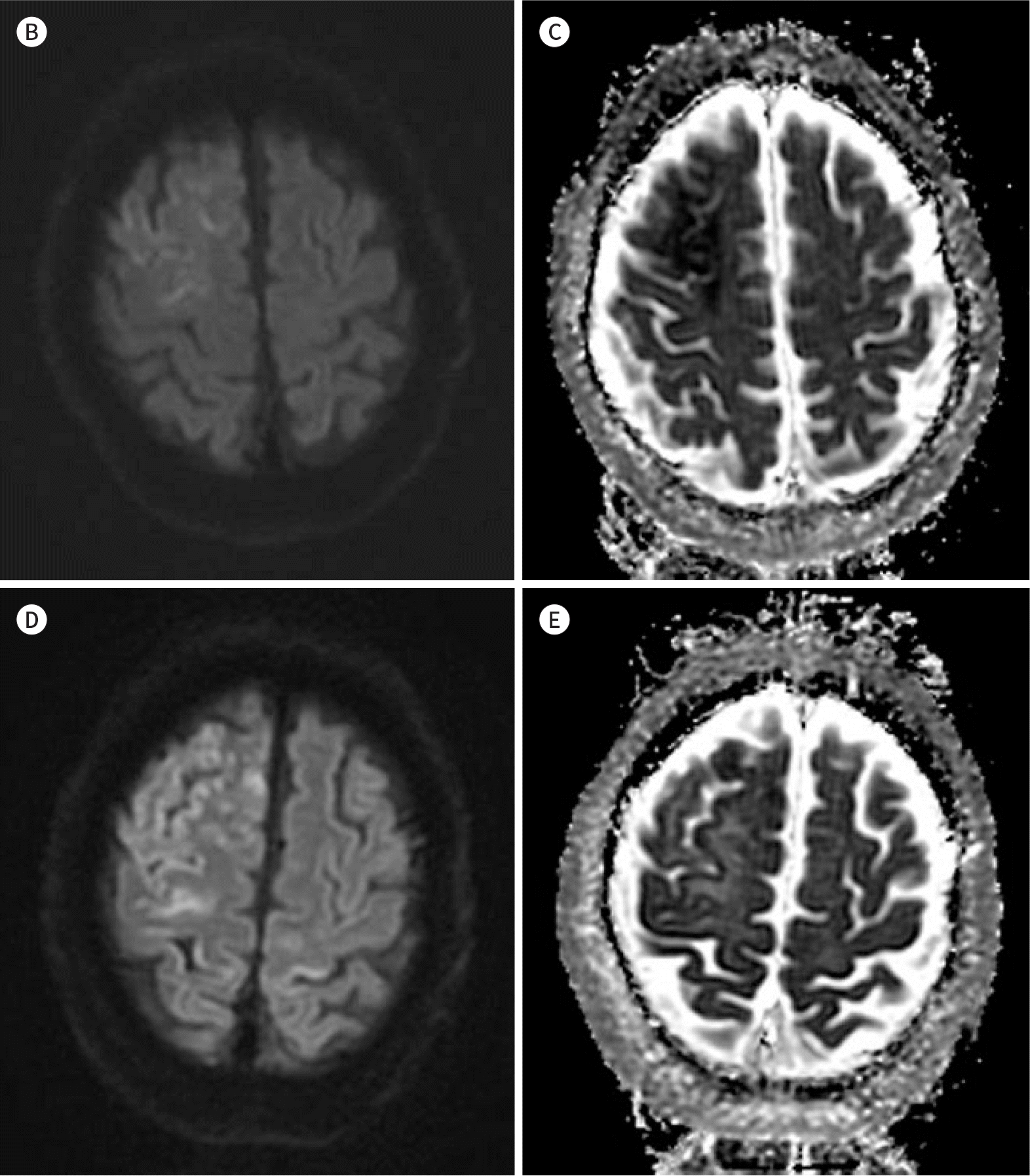
Fig. 1.
A 68-year-old man who developed cerebral air embolism after the removal of a central venous catheter. B, C. Initial brain DWI with a B value of 1000 (B) and an ADC map (C) depicting an acute cerebral infarction in the right frontal lobe. D, E. Short-term follow-up (a day after initial DWI) brain DWI with a B value of 1000 (D) and an ADC map (E) depicting increased extent of acute cerebral infarction in the right frontal lobe. In addition, newly developed acute cerebral infarction can be observed in the left postcentral gyrus. ADC = apparent diffusion coefficient, DWI = diffusion-weighted image
 XML Download