

 PDF
PDF ePub
ePub Citation
 Print
Print


INTRODUCTION
Snaps usually result due to the sudden displacement of an anatomic or pathologic structure during the movement of a neighboring joint (1).
Most cases of snapping knees are caused by intra-articular factors such as meniscal injury, synovial plicae, and loose bodies; however, they are rarely caused by extra-articular factors (2).
Among the extra-articular causes, snapping syndrome in the pes anserinus is rare, with a limited number of cases being described in the literature. In the majority of these cases, snapping occurred either during extension or both flexion and extension movements, and rarely during flexion of the knee alone.
The currently understood mechanism of snapping pes anserinus was first described by Bae and Kwon (3), who proposed impingement of the semitendinosus and gracilis tendons against the tibial condyle at 15° extension from flexion movement of the knee.
The current report presents a case of snapping of the pes anserinus during flexion movement of the knee, diagnosed by dynamic ultrasonography.
CASE REPORT
A 23-year-old male skier presented with complaints of pain and snapping sounds in both knees. The symptoms had been present for six years with no specific history of trauma. Pain in the knees was associated with a palpable “popping” sensation in the posteromedial region during active flexion movements only. Clinical examination did not reveal any angular deformity, limb length discrepancy, or muscle atrophy, and the patient had a normal Q-angle. The popping sound was reproduced on active movement of both the knees from neutral extension to 45° flexion and was associated with a painful sensation. The area of snapping was mildly tender and located at the posteromedial region of the knee. There was no joint effusion, overlying skin erythema, or ecchymosis.
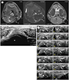
Serial axial MRI of the right knee performed over the years following onset of symptoms did not show any changes in the contour of the flexor tendon, muscle, or bone that explained the snapping (Fig. 1A). No muscular or ligamentous injury was identified on static ultrasonography (Fig. 1B).
Dynamic ultrasonography over the posteromedial region of right knee was performed with the patient lying in the prone position.
With this modality, active flexion and extension of the right knee was observed. Twisting and folding of the gracilis tendon was observed between the sartorius muscle and semitendinosus tendon, on flexion of the knee by 15°. When the patient flexed his knee by 45°, sudden bouncing-off of the gracilis tendon occurred during passage by the sartorius muscle, and a snapping sound was heard. The patient complained of pain until the knee flexed to 45°, but did not experience further pain till the gracilis tendon bounced off and the knee was completely flexed (Fig. 1C, Supplementary Video 1 in the online-only Data Supplement). Sonographic tenderness was also observed in this region, and the patient was diagnosed as a case of snapping pes anserinus.
DISCUSSION
Extra-articular snapping of the knee results from movements of the popliteus, biceps femoris, iliotibial band, or patellar tendons. Snapping pes anserinus syndrome is the main extraarticular cause of medial knee snapping (1).
The patient in the present study experienced bilateral knee pain for six years with no specific findings on serial MRI investigations. However, dynamic ultrasonography allowed accurate diagnosis of the condition as snapping pes anserinus.
The patient was placed in the prone position and the sonographic transducer was placed transversely in the posteromedial region of the right knee. On the monitor, the posteromedial region was superficial, with the sartorius muscle on the left and the semitendinosus tendon on the right side. The gracilis tendon was visible between the sartorius muscle and the semitendinosus tendon, and the medial femoral condyle was deep to this. The patient was then instructed to flex and extend the right knee.
Lyu and Wu. (4) and Bae and Kwon (3) initially described snapping syndrome in the pes anserinus and Bae and Kwon (3) specifically described the snapping mechanism that results from impingement of the semitendinosus and gracilis tendons against the tibial condyle at 15° extension of the knee when returning from a flexed position. The patient in the present study was diagnosed with snapping syndrome in the pes anserinus; however, the snapping occurred only during active flexion of the knee, which is a new mechanism different from that described previously. Twisting and folding of the gracilis tendon occurred during active flexion movement, which suddenly popped out as it passed by the sartorius muscle, causing a snapping sensation.
We propose that the twisting and folding of the gracilis tendon with resulting mechanical friction against the sartorius muscle causes severe pain to the patient. Furthermore, we assert that this pain is caused by a mechanism other than impingement between the semitendinosus and gracilis tendons and the tibial condyle, as proposed initially by Bae and Kwon (3).
Deslandes et al. (5) reported a new mechanism of snapping hip syndrome that occurred in the iliopsoas tendon. The study found that the extra-articular cause of hip snapping was a dynamic interaction between the iliopsoas tendon and the corresponding muscle, rather than impingement between the iliopsoas tendon and iliopectineal eminence or lesser trochanter, as reported in the earlier literature. Similarly, the snapping that occurred at the medial aspect of the knee of the patient in the present study may have been caused by the dynamic interaction between the gracilis tendon and the surrounding muscles in the posteromedial region of the knee. This mechanism is unlike the snapping caused by impingement of the tibial condyle and pes anserinus, as reported in the previous article.
A recent study presented a case similar to that described in the current study. Shapiro et al. (6) reported the case of a 31-year-old female patient with right knee snapping symptom, with no previous history of traumatic injuries and no abnormalities seen on static MRI. The authors in the above-mentioned study also observed that the snapping was secondary to the popping of the gracilis tendon caused by abnormal movement of the tendon against the surrounding muscle, which is similar to the observations of the present case.
The etiology of snapping pes anserinus has been well described in several studies. Snapping pes anserinus is known to result due to exostosis of the underlying bone (78) or a deficiency in the accessory bands that stabilize the pes anserinus (1), in addition to instability of the pes anserinus tendon due to laxity of the ligament following trauma (9), or continuous overloading of the anterior aspect of the knee (310).
Limited studies have reported cases of snapping pes anserinus during flexion movement of the knee alone. Gokhan et al. (7) described the case of a 79-year-old female patient who experienced a snapping sensation only after flexion of the knee after left total knee arthroplasty. Dynamic ultrasonography confirmed that the pes anserinus translocated over a posteromedial tibial osteophyte and the snapping sensation disappeared after excision of the tibial osteophyte. However, in the present case, no abnormal bony lesion such as an osteophyte was observed.
The current study presented a case of snapping pes anserinus in bilateral knees of a 23-year-old skier with no previous history of trauma. No bony or intra-articular abnormality was found on previous static MRIs and static ultrasonography. The posteromedial aspect of the right knee was examined by dynamic ultrasonography with the patient lying in prone position. Unlike the snapping mechanism of the pes anserinus described in previous studies (13), we observed a new mechanism by which the gracilis tendon causes snapping by dynamic interaction with the surrounding muscles only during the flexion of the knee. In addition, as described in multiple articles (14610), this snapping mechanism can be diagnosed accurately with dynamic ultrasonography.
An understanding of the various mechanisms that cause snapping of the pes anserinus, as seen on dynamic ultrasonography, allows accurate diagnosis and treatment of patients with snapping knees.
Supplementary Video Legend
Video 1. Transverse dynamic ultrasonography of the right knee in prone position during active flexion movement. While the patient bends the knee by 15°, twisting and folding of the gracilis tendon is observed. Following this, popping-out of the gracilis tendon is observed as it passed by the sartorius muscle.

 XML Download