

 PDF
PDF ePub
ePub Citation
 Print
Print


INTRODUCTION
In celiac axis stenosis, rich collateral circulations are known to develop from the superior mesenteric artery (SMA) to branches of the celiac artery (1); aneurysm of these collateral vessels has seldom been reported (2). Such aneurysms typically involve pancreaticoduodenal and gastroduodenal arteries, and may rupture spontaneously (3). Here, we report a case in which we performed successful transcatheter arterial embolization for aneurysmal rupture of a previously unreported omental branch, the “gastroepiploico-colic communicating artery.”
CASE REPORT
A 37-year-old male was referred to our emergency department with a complaint of abdominal pain that had been present for 1 day. On physical examination, he complained of tenderness in the right upper abdomen. He had no remarkable history of illness or trauma. His vital signs and hemoglobin level were relatively stable. Blood pressure was 126/76 mm Hg, pulse rate was 81 beats/minute, and hemoglobin level was 13 g/dL.
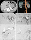
On the CT scan, the celiac stenosis was determined to be a result of median arcuate ligament syndrome. The median arcuate ligament was thickened, thereby compressing the celiac trunk (Fig. 1A). Enhanced abdominal CT and reconstructed CT angiography showed hemoperitoneum with an aneurysm in the right subhepatic area without any other direct extravasation of contrast, so the aneurysm was the convincing focus of bleeding (Fig. 1B). The aneurysm appeared to be located on a branch of the gastroduodenal artery (GDA) or SMA. Because the patient denied any trauma history or coagulopathy, the aneurysm was suspected to have ruptured spontaneously.
After taking CT scan, the hemoglobin level reduced to 11.9 g/dL, and packed red blood cell transfusion was initiated. Additionally, urgent arterial angiography was performed to establish a diagnosis and initiate proper treatment.
Both celiac and SMA angiography procedures were necessary for further evaluation. The GDA could not be visualized on celiac angiography. Only the common hepatic artery, proper hepatic artery (PHA) and splenic artery were shown. (Fig. 1C). However, SMA angiography revealed opacification of the GDA and other celiac branches, including the PHA and splenic artery (Fig. 1D); this was suggestive of celiac stenosis with collateral vessels. This patient had collateral pathways for celiac stenosis, including the pancreaticoduodenal arcades, dorsal pancreatic artery, and the communicating vessel between the right hepatic artery and SMA.
The aneurysm demonstrated on CT was then visualized on SMA angiography; it was in the right subhepatic area, as expected, but was present in an abnormal communicating artery between the right gastroepiploic and right colic arteries (Fig. 1E).
For treatment, embolization was performed at the distal and proximal portions of the aneurysm in the abnormal communicating artery with microcoils (6 mm/2 cm × 8, 8 mm/4 cm × 1, 4 mm/2 cm × 1, Tornado; Cook Medical, Bloomington, IN, USA) by using a microcatheter (Progreat 2.0; Terumo Interventional Systems, Somerset, NJ, USA) (Fig. 1F). Immediate follow-up SMA angiography showed complete embolization of the aneurysm without any immediate complication.
After the embolization, the patient's abdominal pain was relieved and the anemia did not worsen. A CT scan obtained 2 days after the procedure showed improved hemoperitoneum and no evidence of re-bleeding. He was discharged on the eighth hospital day without any additional treatment.
DISCUSSION
Pancreaticoduodenal arcades, dorsal pancreatic artery, the arc of Bühler, and a communicating channel between the right hepatic artery and SMA are known as collateral pathways in celiac stenosis (1). However, to our knowledge, a communicating artery between the gastroepiploic and right colic arteries in celiac stenosis has not been reported. We named this aberrant anastomosis “gastroepiploico-colic communicating artery.” It may be a collateral pathway arose on the omental anastomosis between the branches of gastroepiploic artery and colic arteries. We speculate that increased blood flow from the SMA to the celiac branches led to reopening of this communicating channel. Aneurysms and ruptures of collateral arteries in celiac trunk stenosis have been reported (4). Common affected arteries are pancreaticoduodenal artery and GDA (3). In our case, there was an aneurysm in the gastroepiploico-colic communicating artery. An aneurysm typically develops in the weakest vessel; in our patient, this was the gastroepiploico-colic communicating artery, which was newly opened. Otherwise there could be other etiologies that the patient didn't recognize, such as minor trauma.
Transcatheter arterial embolization is now the treatment of choice for vascular aneurysm and aneurysmal rupture. Yet, the timing of preventive treatment for unruptured aneurysm is still controversial. According to Ogino et al. (4), embolization is necessary for all discovered aneurysm in the splanchnic artery because of its high frequency of rupture. However, some authors believed that the relative size of the aneurysm to the originating vessel is the most important factor and the bleeding occurred in aneurysms which had aneurysm-to-artery ratio of at least 3 in their study (5). Also, others suggested that the indication of interventional treatment is visceral aneurysm with 2 cm or more in diameter (6).
Lastly, It is important that in a patient with celiac stenosis, vascular evaluation is needed to screen for aneurysm since aneurysmal rupture of splanchnic vessels can be life-threatening.
 XML Download