

 PDF
PDF ePub
ePub Citation
 Print
Print


INTRODUCTION
Osteomas, which are benign neoplasms consisting of mature osseous tissue, are uncommon in the soft tissues of the head and neck (1); however, when osteomas do occur in these body parts, they are usually found in certain regions of the head and neck, especially in the localized regions of the paranasal sinuses, maxilla, and mandible (2). Notably, an osteoma of the tongue (lingual osteoma) is rare, and fewer than 100 cases have been reported to date in previous literature. Typically, a lingual osteoma is located in the posterior one-third of the tongue, adjacent to the foramen cecum. It usually occurs asymptomatically in women between 30 and 50 years of age. If symptomatic, patients commonly complain of swallowing difficulties (3).
Owing to the characteristic image findings on CT, the CT scan could be helpful for the diagnosis of lingual osteoma, despite its rarity. Herein, we report a case of lingual osteoma and its CT findings with a review of the existing literature.
CASE REPORT
This report was approved by the Institutional Review Board of Chung Ang University Hospital, and consent was obtained from the patient herself. Ethical approval was not needed as the case report was a retrospective observation of the patient.
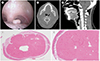
A 39-year-old woman visited our outpatient clinic for the evaluation of a mass at the base of her tongue, which was incidentally found approximately one and a half years ago. She complained of a recently developed lump in her throat. Laboratory studies revealed no abnormalities. Laryngoscopy showed a small, whitish submucosal mass protruding from the dorsum of the tongue base, without tenderness, and a well-preserved overlying mucosa (Fig. 1A).
Contrast-enhanced CT of the neck showed a well-circumscribed, hyperdense, oval mass arising from the base of the tongue without any enhancing soft tissue component (Fig. 1B, C). The size of the mass was 0.7 × 0.5 × 0.7 cm, and the mean density was 1152 Hounsfield units, similar to that of the cortex of the mandible. There was a thin mucosal lining above the mass, which suggested a submucosal location. No significantly enlarged lymph nodes were found in the head and neck. Initially, the differential diagnosis of the lesion included calcified lingual thyroid and dystrophic calcification based on the CT appearance of the mass on the tongue.
During excisional biopsy under general anesthesia, the location of the mass was thought to be at the base of the tongue, off the midline to the left side at the palatine tonsil level. Grossly, there were no remaining masses after the surgical excision. Microscopically, the mass was composed of a dense, mature lamellar bone (Fig. 1D, E), which was covered with the normal squamous epithelium of the tongue, and was suggestive of a lingual osteoma.
DISCUSSION
Lingual osteoma is a rare and benign tumor typically arising from the tongue base and notably, with less than 100 reported cases (4) and only a limited number of image findings (5). Most patients are in the third or fourth decade of life, and there is a four-fold female predilection (male to female ratio of 1:4). Approximately 60% of the patients are symptomatic, and a sense of a “lump in the throat,” dysphagia, gagging, and nausea are commonly reported (3). There are no correlations between the symptoms and size of the tumor (6). Surgery is the primary treatment for the symptomatic cases, and no recurrences have been reported (7).
There are a few hypotheses on the etiology of lingual osteoma, mainly derived from the characteristic location of the lesion. One theory is that the pluripotent mesenchymal cells from the first and third branchial arches, which meet in the foramen cecum, can facilitate the development of lingual osteoma (2). Another theory is that lingual osteoma may develop from reactive or post-traumatic changes (2). However, the osseous lesions caused by trauma-induced chronic inflammation usually do not develop into normal bone structures such as the Haversian system or the fatty marrow. Moreover, a lingual osteoma is composed of well-developed mature bone, except in two previously noted cases (3). Lastly, another theory is that the undescended thyroid remnants may cause abnormal bony proliferation of the tongue (1). Not only lingual osteoma, but also lingual thyroid remnants and embryologically displaced intralaryngeal thyroid tissue show a female predilection. Moreover, metaplastic ossification of thyroid tissue is also reported in colloid goiters and thyroid cysts (1). Therefore, lingual osteomas can be considered as the derivatives of thyroid remnants.
On review of the CT findings, the lingual osteoma was noted as a homogenously hyperdense lesion without enhancement, located at the base of the tongue (5). The margin of the lesion was well-circumscribed, and the attenuation of the lesion was similar to that of the bone. Owing to its typical imaging features, CT could be helpful in differentiating lingual osteoma from other submucosal tumors of the tongue base. Initial differential diagnosis included calcified lingual thyroid and dystrophic calcification. In case of a calcified lingual thyroid, thyroid tissue usually shows contrast enhancement on the midline of the tongue base (8). In our case, the lesion was composed of hyperdense bony tissue without any enhancing soft tissue. Dystrophic calcification rarely appears in the injured tissue or at the site of radiation therapy in the head and neck region (9). Our patient had neither a specific medical history nor history of injury. Accordingly, the former two differential diagnoses could be ruled out. Other soft tissue-attenuated tumors near the foramen cecum, such as tonsillar hypertrophy, lingual thyroid, salivary gland tumor, and squamous cell carcinoma, should be ruled out using CT images (1). Furthermore, it can be distinguished from an osteosarcoma with local bone destruction or superficial ulcer, or from osteolipoma with soft tissue density mixed with bone density (35).
The MRI findings of lingual osteoma have also been reported to show a mass with significantly low signal in both T1-weighted and T2-weighted images because of its heavily calcified mature bone structure (1). In contrast, the lingual thyroid, which has an anatomically similar location, can be distinguished by the intermediate signal intensity and the slightly high signal intensity on the T1-weighted and T2-weighted images as normal thyroid, respectively (3).
CT is superior in detecting calcifications in the head and neck area (10). Since lingual osteoma predominantly consists of mature lamellar bony tissue (1), CT is a more suitable imaging modality than MRI for the preoperative imaging of lingual osteoma. Due to the absence of signal from the calcification and cortical bone on MRI, all MRI scans should be preceded by a corresponding CT and radiograph. Therefore, CT would be a better imaging modality than MRI to diagnose a lingual osteoma.
In this case study, we demonstrated the characteristic CT findings of a surgically verified lingual osteoma. It is a well-circumscribed, hyperdense submucosal mass, which appears near the foramen cecum. Although the tumor is rare, knowing the typical CT findings of a lingual osteoma could be helpful for its diagnosis and prevent further unnecessary work-up.
 XML Download