

 PDF
PDF ePub
ePub Citation
 Print
Print


INTRODUCTION
Osteoradionecrosis of the thoracic wall is rare; nevertheless, severe complications of radiation therapy (RT) can occur in patients with breast and lung cancers. Blood vessels that supply the bone with nutrients can be damaged by RT. Vascular compromise with obliterative endarteritis and damage to osteoblasts and osteoclasts is a possible cause of osteoradionecrosis (1). It usually occurs > 1 year after completion of RT in patients treated with doses > 6 Gy (1). Radiological findings of osteoradionecrosis include focal lucent area, periostitis, sclerosis, cortical thinning and irregularity, fatty marrow changes, and insufficiency fractures. The differentiation of osteoradionecrosis from bone metastasis is very important for radiologists to plan appropriate treatment. This case report was approved by the author's Institutional Review Board, and the requirement for informed consent was waived.
CASE REPORT
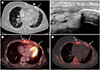
A 65-year-old woman presented with an open wound and pain in her left chest wall. She underwent left radical mastectomy for breast cancer 17 years previously. She subsequently underwent adjuvant chest wall RT with 5000 cGy in 25 fractions, and adjuvant chemotherapy with 8 cycles of 5-florourasil, epirubicin, and cyclophosphamide after surgery. Subsequently, tamoxifen treatment of 20 mg/day for 5 years was continued. The wound on her left chest wall was developed a few days previous to presentation. She denied a history of trauma to her left chest wall. On physical examination, an ulcer approximately 1 cm in size was observed on the left mastectomy site. CT of the chest revealed fractures of the left third and fourth ribs and the sternal body, with severe lytic and sclerotic changes, and cortical irregularity (Fig. 1A). Ultrasonography of the left chest wall revealed diffuse thickening of the skin and increased echogenicity and edematous changes in the subcutaneous fat layer (Fig. 1B). PET/CT of the chest wall demonstrated increased uptake in the sternal body, left ribs, and overlying soft tissue (Fig. 1C). Thus, osteomyelitis with associated cellulitis was initially suspected. However, bone metastasis could not be excluded based on these imaging findings. A skin punch biopsy of the ulcer lesion at the left chest wall was performed and revealed fibrinoid materials and calcifications without viable cells. Bone biopsy was recommended to differentiate it from skeletal metastasis, however, the patient refused to undergo the procedure. Conservative treatment with pain control and management of the open wound was implemented. Two years later, follow-up PET/CT was performed and continued to demonstrate increased uptake of radioisotope in the sternal body and ribs, with progressed sclerotic changes and non-union state of fractures (Fig. 1D). Therefore, the possibility of radiation-related osteonecrosis was raised. Conservative treatment, including analgesia, was performed for the patient. Over the 5-year follow-up period, the patient's anterior rib pain resolved; however, her wound persisted with repeated improvement and deterioration.
DISCUSSION
The primary goal of RT is the eradication of microscopic residual disease adjacent to the tumor site and to eliminate the possibility of multicentric disease. Usually, external beam radiation is used for the treatment of breast cancer; it affects the adjacent structures as well as the original site of the tumor. Several complications associated with RT may occur. Early complications (during the weeks to months after completion of RT) include skin changes, breast edema, dystrophic calcifications, fat necrosis, radiation-induced pneumonia and pleural effusion. Months to years after the completion of RT, fibrotic changes in the breast(s), glandular atrophy, lactational difficulty, lymphedema, brachial plexopathy, bone fracture, pulmonary fibrosis and pericardial disease can occur (1). Late complications (> 10 years after completion of RT) include cardiomyopathy and secondary malignancies (12). Radiation-induced rib fracture is a very rare, late complication of conventionally fractionated RT for breast and lung cancer. The previously reported incidence is between 0.1% and 5% (23). Severe osteoradionecrosis of the ribs is extremely rare, with only a few case reports published over the past decades (45). While osteoradionecrosis of the ribs is rare, the mandible is a relatively common site of osteoradionecrosis due to the higher radiation doses administered for head and neck cancers, and the poor vascular system of mandible (6).
Radiologically, osteoradionecrosis may present as focal lucent area in bone, periostitis, sclerosis, cortical thinning and irregularity, fatty marrow change, and insufficiency fractures (2). Bone scan will demonstrate decreased uptake of radioisotope in the early stages and, later, increased uptake of radioisotope with fracture will appear. On PET scan, osteoradionecrosis may appear as a false-positive finding. Multiple previous studies have reported the relative unreliability of increased uptake on PET/CT in differentiating osteonecrosis from skeletal metastasis (7). Skeletal metastasis usually demonstrates higher uptake compared with osteonecrosis; however, there is significant overlap. Some CT findings are helpful for differentiating bone metastasis from osteoradionecrosis. For example, a discrete, associated solid or cystic mass is a significant diagnostic indication for the presence of skeletal metastasis (7). In one previous study, a permeative pattern of bone loss (< 75% loss of total bone trabecula) was more commonly seen in osteoradionecrosis, and lucent pattern (> 75% loss of total bone trabecula) is more often observed in metastasis (7). Because, irradiation leads to relatively hypoxic, hypocellular, hypovascular substrate with an inconsistent ability to remodel tissue loss, this would lead to relatively less bony loss than would be evident when tissue is being actively destroyed and replaced by tumor. Additionally, intraosseous gas may be evident in osteoradionecrosis because of superimposed osteomyelitis, but not in metastasis (7).
Irradiated bone is susceptible to infection and is associated with a high risk for bone sarcomas. The differentiation of osteoradionecrosis from bone metastasis is very important for radiologists to plan appropriate treatment. Predisposing factors for osteoradionecrosis include trauma, infection, inflammation, overdose radiation, bony invasion by tumor, tumor location around the bone, and individual sensitivity of the patient (8). The most frequent symptoms of osteoradionecrosis include pain, infection, and pathological fractures. Osteoradionecrosis can be diagnosed through joint evaluation of clinical findings, pathology, and radiology (5). Biologically, radiation-induced fibroatrophic changes, chronic inflammation, and tumor necrosis factor-α have been suggested to be potential causes of osteoradionecrosis (9). Various treatments such as pentoxifylline, vitamin E and clodronate for inhibiting tumor necrosis factor-α have been developed (10). Hyperbaric oxygen has been proposed as an adjunctive therapy and may improve outcomes (8). Surgical debridement with reconstruction of the chest wall can be an option for patients who fail treatment with conservative management (5). However, there are no established treatment guidelines for osteoradionecrosis. The management of radiation-induced rib fracture is to optimize analgesia, exclude recurrent disease, and trialling established treatments for osteoradionecrosis (5).
In summary, we reported a case of osteoradionecrosis affecting the left ribs following adjuvant chest wall RT, and presented a brief review of the literature. Radiologists should be aware of imaging findings of osteoradionecrosis to differentiate it from bony metastasis.
 XML Download