Journal List > Anat Cell Biol > v.52(3) > 1134438

Iwanaga, Wong, Kikuta, and Tubbs: Stafne bone cavity: a rare cadaveric case report

Abstract

The Stafne bone cavity (SBC), also called the static bone cavity, salivary inclusion cyst, latent cyst, and lingual bone defect is an asymptomatic bony defect that is commonly located inferior to the mandibular canal and slightly above the inferior border of the mandible. It is rare to see the actual bony defect in the cadaver because of its relatively low incidence of 0.1% to 6.06%. We report a unilateral SBC found in a 76-year-old at death male Caucasian cadaver and involving the right mandible. The SBC was oval in shape with a smooth surface and measured 10.8×6.0 mm. The SBC was continuous with the right mylohyoid groove. Since actual photographs of the SBC are lacking in the literature, this report might provide additional insight for better understanding the SBC.

Introduction

The Stafne bone cavity (SBC), first reported by Edward Stafne in 1942 [1], also called the static bone cavity, salivary inclusion cyst [2], latent cyst [3], and lingual bone defect [4], is an asymptomatic bony defect that is commonly located inferior to the mandibular canal and slightly above the inferior border of the mandible [15]. The reported incidence of the SBC ranges from 0.1% to 6.06% [56]. It is usually found on one side but occasionally, on both sides of the mandible [7]. The submandibular gland, fat tissue, connective tissue, lymphatic tissue, muscle or vessels have all been considered as potential contents of the SBC [4]. In the clinical setting, most SBC is found incidentally during panoramic radiography. Since the SBC is an asymptomatic bone cavity and does not require treatment, it is relatively rare to see the actual bony defect without radiographic images. Therefore, herein, we report a case of unilateral SBC in a cadaver.

Case Report

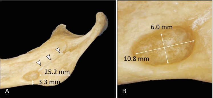
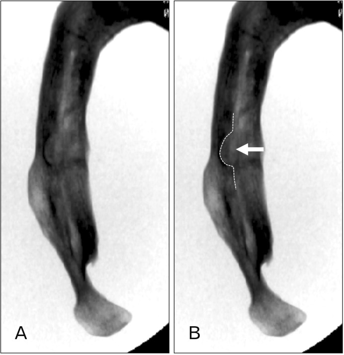
A 76-year-old at death male Caucasian cadaveric head (fresh-frozen) was dissected. During routine dissection of the head, the mandible was dissected, and the soft tissues of the mandible were removed. After the mandible was separated from the head, a large bony defect was found on the lingual surface of the right mandible, which was located 25.2 mm anterior to the angle of the mandible and 3.3 mm superior to the inferior border of the mandible. The shape and size of the bony defect was oval and 10.8×6.0 mm, respectively (Figs. 1, 2). The bony defect had a smooth surface and did not show any pathological condition. Therefore, it was determined to be an SBC. The SBC had continuity with the right mylohyoid groove. The depth of the SBC and width of the mandible at the SBC was 4.15 mm, 3.14 mm, respectively. The SBC was not observed on the left side.

Discussion

Studies on the SBC using dry mandibles were reported by Harvey and Noble in 1968 [7] and Kay in 1974 [8]. Since then, most of the reported studies examined either patients' panoramic radiography or computed tomography. The etiology of SBC is unclear. Many hypotheses exist regarding the cause of the bony defect, including a hypertrophic lobe of the submandibular gland, erosion by vascular compression [9], or incomplete Meckel cartilage ossification [9]. Minowa et al. [9] hypothesized that an SBC results from bone resorption or an overlying lipoma. In a case report by Kaya et al. [10], an ectopic parotid gland was found in the SBC. SBCs are most frequently found in patients over 50 years old [9] and there have been no reports of SBCs in patients younger than 10 years old [11]. On conventional panoramic radiographs, the SBC usually resembles a unilocular cystic lesion with well-defined borders. Multiple SBC was also reported by Aguiar et al. [12].
Histological examination by Harvey and Noble [7] revealed such bony defects did not invade the mandibular canal. However, the most recent study by Hisatomi et al. [13] demonstrated that an SBC in the premolar area might be misdiagnosed as a periapical lesion or odontogenic cyst. Thus, an SBC is occasionally not located inferior to the mandibular canal. Barker [14] also reported a case with an SBC situated on ascending ramus. An anthropological study conducted by Wasterlain and Silva [15] investigated ancient mandibles with an SBC. These SBC were most commonly situated below the third molar or inferior to the mylohyoid groove. In the present case, the mylohyoid groove had continuity with the SBC.
Even though the SBC is asymptomatic and does not require treatment, this anomaly should be taken into consideration by dentists and oral surgeon in order to avoid misdiagnosis.

Notes

Author Contributions:

  • Conceptualization: JI, RST.

  • Data acquisition: JI, TLW, SK.

  • Data analysis or interpretation: JI, TLW, SK.

  • Drafting of the manuscript: JI, TLW, SK.

  • Approval of the final version of the manuscript: all authors.

Conflicts of Interest: No potential conflict of interest relevant to this article was reported.

References

1. Stafne EC. Bone cavities situated near the angle of the mandible. J Am Dent Assoc. 1942; 29:1969–1972.
2. Fordyce GL. The probable nature of so-called latent haemor rhagic cysts of the mandible. Br Dent J. 1956; 101:40–42.
3. Rushton MA. Solitary bone cysts in the mandible. Br Dent J. 1946; 81:37–49. PMID: 20992458.
4. Etoz M, Etoz OA, Sahman H, Sekerci AE, Polat HB. An unusual case of multilocular Stafne bone cavity. Dentomaxillofac Radiol. 2012; 41:75–78. PMID: 22184629.
5. Quesada-Gómez C, Valmaseda-Castellón E, Berini-Aytés L, Gay-Escoda C. Stafne bone cavity: a retrospective study of 11 cases. Med Oral Patol Oral Cir Bucal. 2006; 11:E277–E280. PMID: 16648768.
6. Li B, Long X, Cheng Y, Wang S. Cone beam CT sialography of Stafne bone cavity. Dentomaxillofac Radiol. 2011; 40:519–523. PMID: 22065802.
7. Harvey W, Noble HW. Defects on the lngual surface of the mandible near the angle. Br J Oral Surg. 1968; 6:75–83. PMID: 5246825.
8. Kay LW. Some anthropologic investigations of interest to oral surgeons. Int J Oral Surg. 1974; 3:363–379. PMID: 4217312.
9. Minowa K, Inoue N, Sawamura T, Matsuda A, Totsuka Y, Nakamura M. Evaluation of static bone cavities with CT and MRI. Dentomaxillofac Radiol. 2003; 32:2–7. PMID: 12820846.
10. Kaya M, Ugur KS, Dagli E, Kurtaran H, Gunduz M. Stafne bone cavity containing ectopic parotid gland. Braz J Otorhinolaryngol. 2018; 84:669–672. PMID: 27133906.
11. Hansson LG. Development of a lingual mandibular bone cavity in an 11-year-old boy. Oral Surg Oral Med Oral Pathol. 1980; 49:376–378. PMID: 6928587.
12. Aguiar LB, Neves FS, Bastos LC, Crusoé-Rebello I, Ambrosano GM, Campos PS. Multiple stafne bone defects: a rare entity. ISRN Dent. 2011; 2011:792145. PMID: 21991486.
13. Hisatomi M, Munhoz L, Asaumi J, Arita ES. Stafne bone defects radiographic features in panoramic radiographs: assessment of 91 cases. Med Oral Patol Oral Cir Bucal. 2019; 24:e12–e19. PMID: 30573717.
14. Barker GR. A radiolucency of the ascending ramus of the mandible associated with invested parotid salivary gland material and analogous with a Stafne bone cavity. Br J Oral Maxillofac Surg. 1988; 26:81–84. PMID: 3422827.
15. Wasterlain SN, Silva AM. Study of stafne's defects in Late Neolithic, Late Roman, Medieval and Modern skeletal samples from Portugal. Int J Osteoarchaeol. 2012; 22:423–434.
Fig. 1

Stafne bone cavity (SBC) of the right mandible. (A) SBC located 25.2 mm anterior to the posterior border of the ramus and 3.3 mm superior to the inferior border of the mandible. Note the mylohyoid groove (arrowheads) has continuity with the SBC. (B) Magnified photo of Fig. 1A.

acb-52-354-g001
Fig. 2

Fluoroscopy of the right mandible (superior-inferior view) showing depth of Stafne bone defect. (A) Plain radiography. (B) Pointing the Stafne bone defect (dotted line and arrow).

acb-52-354-g002
TOOLS
Similar articles