

 PDF
PDF ePub
ePub Citation
 Print
Print


Introduction
Arterial variations in upper limb are commonly reported. Rodríguez-Niedenführ et al. (2001) [1] conducted a metanalytic study with a large sample of the upper limbs and categorized the arterial variations based on topographical criteria into 21 types, including a classical description of upper limb arteries. Superficial arterial variations accounting for 4.2% of all arterial variations are hazardous during any invasive procedures of the upper limb, from routine intravenous injections to surgeries. Recent studies show that these arterial variations are usually associated with muscular variations, the most common being the absent or inverted palmaris longus [2]. Palmaris profundus, a rare anomalous variation of palmaris longus has been reported in carpal tunnel syndrome as its tendon was associated with median nerve in the carpal tunnel [3]. This study reports a unique variation in the upper limb arterial pattern—the presence of bilateral superficial brachioulnar artery (SBUA) associated with palmaris profundus arising from the deeper aspect of a fleshy palmaris longus on right side and an abnormal radicle of musculocutaneous nerve to the median nerve on the left side.
Case Report
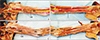
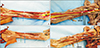
During the routine upper limb dissection for undergraduate medical students, the authors spotted an unusual constellation of upper limb arterial, muscular and nervous variation in a 65-year-old female of Indian origin. In the present case, SBUA was noticed bilaterally. On the left side, SBUA was originating from the third part of axillary artery proximal to the origin of anterior and posterior circumflex humeral arteries; on the right side, SBUA was originating from brachial artery just below the origin of profunda brachii artery and above the level of insertion of coracobrachialis (Figs. 1, 2). In the arm bilaterally, the SBUA was running medial to the normal brachial artery but in a more superficial plane. In the cubital fossa, SBUA was superficial to the pronator teres muscle and continued as the superficial ulnar artery on both sides. In the forearm, SBUA continued its medial course superficial to the flexor muscles. In the distal part of the forearm, SBUA passed deep to the tendon of palmaris longus and palmaris profundus and then was medial to the palmaris longus tendon in the wrist. Its contribution to the superficial palmar arch was similar to that of the normal brachial artery bilaterally.
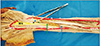
On the right side, palmaris longus also had a rare variation of a fleshy belly instead of a long tendon (Figs. 1, 2). Authors noticed Palmaris profundus originating from the posterior aspect of the epimysium of the palmaris longus and a few of its fibers arising from the common flexor origin. As the name implies, palmaris profundus belly was deep to palmaris longus, and the tendon of palmaris profundus passed deep to flexor retinaculum and was then inserted into the under surface of the palmar aponeurosis (Fig. 3). In the carpal tunnel, it was accompanied by the median nerve. Palmaris profundus received nerve supply from the anterior interosseous nerve. Palmaris longus in the left side had the characteristic long tendon. In the left side, a few fibers of musculocutaneous nerve joined the median nerve below the level of the insertion of the coracobrachialis. Bilaterally, the brachial artery divided into radial artery and common interosseous branch at the level of the neck of the radius. Further course of radial and common interosseous arteries was normal.
Discussion
The subclavian artery continues as axillary artery at the posterior margin of the first rib, which in turn continues as brachial artery at the inferior margin of teres major. Usually, the brachial artery bifurcates into the ulnar and radial artery at the level of the neck of the radius. The ulnar artery is usually larger in diameter than the radial artery and gives rise to the common interosseous artery. The ulnar artery runs with the ulnar nerve between the superficial and deep flexors. At the wrist, the ulnar artery passes superficial to flexor retinaculum. In the palm, ulnar artery contributes to superficial and deep palmar arch [4].
When there is an additional brachial artery, it may be either accessory or superficial brachial artery. Accessory brachial artery joins with a normally situated brachial artery in the arm itself, while the superficial brachial artery runs superficial to the normal brachial artery and usually continues as the radial artery or the ulnar artery. The superficial brachial artery may arise from brachial artery in mid-arm level or rarely from the axillary artery and will be superficial and medial to the median nerve. When the ulnar artery runs superficial to flexor muscles, it is termed as a superficial ulnar artery. In few cases, these superficial brachial arteries may continue as the superficial ulnar or superficial radial artery and are known as superficial brachioulnar or superficial brachioradial artery, respectively. In such variations, the common interosseous artery may arise either from radial artery or directly from brachial artery [1].
In the present case, the superficial brachial artery arises from the third part of the axillary artery on the left side and on the right, it arises from brachial artery itself (Fig. 2). The typical superficial ulnar artery runs superficial to all the forearm muscles, but in the present case, SBUA runs superficial to all the flexors except palmaris longus bilaterally. A similar course of the SBUA has been reported in three studies previously [5678]. These variations result from persistence and enlargement of initial capillary plexus which regresses during the usual course of development of the upper limb arterial system [1].
Arterial system of the upper limb develops in 5 stages. In stage 1 and 2, axial system vessels include the axillary artery, the brachial artery, the anterior interosseous artery and the median artery are developed. In stage 3, the ulnar artery sprouts from brachial artery. Then a superficial brachial artery develops from the axillary artery and continues as the radial artery in stage 4. Later in stage 5, regression of the median artery and regression and anastomosis of superficial brachial artery with brachial artery gives adult pattern of radial artery arising from brachial artery. In the current scenario, the ulnar artery might develop the same as radial artery development and persist as SBUA due to high hemodynamic predominance of the superficial system over the deep system.
Usually, SBUA is associated with absent or inverted palmaris longus [29]. The long fleshy palmaris longus was reported as a very rare variant, which may be associated with carpal tunnel syndrome [10]. In the present case, an SBUA on the right side was associated with long fleshy palmaris longus and palmaris profundus. To the best of our knowledge, no such association has been reported previously.
Palmaris profundus, a rare anomalous muscle was first reported in 1908 [3]. Reimann et al. [11] reported only 1 case out of 1,600 cadaveric limbs. Several variants of palmaris profundus have been reported in the literature [3]. Palmaris profundus has been classified based on its origin into subtypes: (1) from the proximal or mid-third of the radius (flexor carpi radialis profundus muscle [type 1], or palmaris profundus radial muscle if bicipital origin [type 3]); (2) from the fascia of the flexor digitorum superficialis (palmaris profundus longus muscle [type 2]); and (3) from the anterior surface of the distal ulna (palmaris profundus ulnaris muscle [type 4]). The innervation is from the anterior interosseous nerve in types 1, 2, and 3 or from the ulnar nerve in type 4. In addition to these origins, Parmaris profundus may arise from the common flexor origin, palmaris longus or the epimysium of the flexor pollicis longus. In the present case, palmaris profundus arises from the undersurface of epimysium of palmaris longus and common flexor origin, which is a rare variant of the same.
Abnormal radicle between the musculocutaneous nerve and median nerve are very well reported in the literature [12]; here we noticed the abnormal communication between musculocutaneous nerve and median nerve below the level of insertion of coracobrachialis. Association of SBUA with variation in nerve distribution of upper limb has been rarely reported.
The presence of the SBUA is lethal as it is vulnerable to injury during interventions in the upper limb from intravenous injections to plastic surgery [1]. Superficial arteries may be mistaken as veins and any intravenous injections into these may lead to ischemia due to blockage of capillaries followed by loss of limb function distal to the site of injection [1]. If the palmar arch is incomplete, damage to these superficial vessels during surgical procedures may lead to ischemia of hand. Carpal tunnel syndrome is more common in the presence of palmaris profundus. SBUA with palmaris profundus is more prone to damage during upper limb surgeries.
The present study reports a rare association of SBUA with palmaris profundus which may have significant clinical fallout. Arterial variations in upper limbs are very common. Thorough knowledge about these variations is necessary to avoid iatrogenic injuries during carpal tunnel release or any other upper limb surgery. The absence of palmaris longus can be detected by a simple clinical test [13], that helps the surgeon to look for underlying vascular anomalies, but to detect the presence of palmaris profundus requires a thorough knowledge about possible muscular variations and radiological imaging like ultrasound and MRI.
 XML Download