

 PDF
PDF ePub
ePub Citation
 Print
Print


Introduction
Coronary arteries are the branches of ascending aorta and they supply the myocardium of the heart. Some of the variations like, absence of one of the coronary arteries [1], origin from pulmonary trunk [2], and origin of both coronary arteries as a common trunk [3] have been reported in the literature. The anatomical variations may or may not affect the normal functioning of the heart. But they may cause problems in the radiological procedures like cannulations. We report a very rare variation that could cause problems in radiological procedures related to the coronary arteries.
Case Report
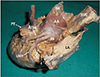
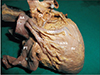
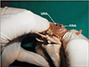
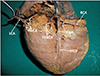
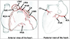
During dissection classes for undergraduate medical students, we observed the absence of left coronary artery in an adult male cadaver aged 78 years, who had a natural death due to old age. The left aortic sinus was dilated and it gave origin to anterior interventricular and left marginal arteries independently (Fig. 1). The left marginal artery was huge and the circumflex artery arose from it at a distance of 2 cm from the left aortic sinus (Fig. 2). When the ascending aorta was opened, we could see two separate openings, one each for the anterior interventricular and left marginal arteries (Fig. 3). The diameters of both openings were approximately 2 mm. The distribution of the anterior interventricular artery was as described in the textbooks of anatomy except that it supplied major part of interventricular artery as the posterior interventricular artery was small. The left marginal artery supplied major part of the left and posterior (inferior) surfaces of the left ventricle. The circumflex artery had a normal course, and termination but it supplied a smaller part of left ventricle than usual. It terminated on the posterior aspect of the heart by giving a few branches, which entered the myocardium (Fig. 4). The right coronary artery had a normal origin, course and branching pattern. However, its posterior interventricular branch was small. Posterior interventricular artery terminated in the upper part of the posterior interventricular sulcus. Due to this, the lower part of interventricular septum was entirely supplied by the septal branches of the anterior interventricular branches traveling through its myocardium. There was no direct anastomosis between right and left coronary arteries on the posterior surface of the heart (Fig. 4). No variations were found in the walls and chambers of the heart. A simplified schematic diagram of the variations has been given as Fig. 5.
Discussion
The left coronary artery usually arises from the left posterior aortic sinus and after a short course, divides into anterior interventricular and circumflex arteries. Its possible variations have been well documented. In rare cases, it could be absent congenitally, in which case, the right coronary artery is super dominant [1]. A recent study from Thailand by Kultida et al. (2018) [4] has reported the absence of the left main coronary artery and left circumflex artery in 0.4% cases. Cortes et al. (2018) [5] have recently reported a case of congenital absence of left coronary artery, causing a sudden cardiac arrest during indoor exercise. A case of congenital absence of left circumflex artery with the presence of a super dominant right coronary artery has also been reported [6]. Saglam et al. (2017) [7] have presented a case of single right coronary artery with the absence of left coronary, anterior interventricular, and circumflex arteries. Iliev et al. (2018) [8] have reported the common trunk origin of left coronary artery with a super dominant right coronary artery. The left circumflex artery was hypoplastic [8]. Coşansu et al. (2018) [9], have reported the presence of a twin circumflex artery in a symptomatic patient.
Although many variations of left coronary artery have been reported, origin of anterior interventricular artery and left marginal artery directly from the left posterior aortic sinus have not been reported hitherto. The unique feature of this case is origin of left circumflex artery from the left marginal artery due to the large size of left marginal artery. This anomaly might not cause functional problems as such because both ostia of these vessels were adequately large and all areas of heart were supplied by branches of these vessels. Knowledge of variations of coronary arteries is important during coronary stent placing and bypass surgeries. The current variation might cause difficulties in catheterization of anterior interventricular and circumflex arteries.
 XML Download