

 PDF
PDF ePub
ePub Citation
 Print
Print


Introduction
The human tentorium cerebelli is a crescentic fold of dura mater, situate above the posterior cranial fossa, separating the occipital and temporal cerebral hemisphere from the cerebellum and infra-tentorial part of brainstem. The tentorium cerebelli is a differentiating landmark and divides the cranial cavity into the supratentorial and infratentorial spaces [1]. The tentorium cerebelli is more developed in humans than subhuman species, which may reflect its role in supporting the heavier cerebral hemispheres in the humans [2].
The falx cerebelli is a small sickle-shaped fold of dura mater located below the tentorium cerebelli. Occipital sinus is usually placed along its posterior attachment to the internal occipital crest [3]. Variations in the morphology of the falx cerebelli are very rare. Reported variations include its complete absence, duplication, triplication, fenestration, and variation in dimensions.
The dura mater and its partitions are noticed as early as the 14th gestational week [4]. The developing nervous system induces the formation of the dura mater from adjacent mesenchymal cells [5]. As the development of the dural venous sinuses goes hand in hand with the development of the dural folds, any changes in the morphology of the dural folds may possibly be related with variations of the venous sinuses too.
Case Report
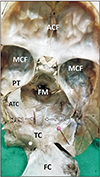
During routine dissection classes for medical students, a concurrent variation of tentorium cerebelli and falx cerebelli was noted in an adult male cadaver aged approximately 75 years. While removing the brain, the falx cerebri was detached from the crista gali and reflected backwards. Initially, the tentorium cerebelli appeared to be normal, with its usual attachments to anterior clinoid process, the superior border of petrous temporal bone and the lips of transverse sulcus. The tentorium cerebelli was detached from the upper border of the petrous part of the temporal bone and retracted backwards along with falx cerebri. At this stage, a partial duplication of tentorium cerebelli was observed. Below the left half of the tentorium cerebelli and above the left cerebellar hemisphere, there was a second fold of dura mater. This fold was about 3cm broad and 5 cm wide and extended from the lower lip of left transverse sulcus behind, to the upper border of the left petrous temporal in front. This fold was made up of meningeal layer of dura mater and did not have any dural venous sinus in it. The accessory tentorium cerebelli was slightly thinner than the main tentorium cerebelli. While lifting the cerebellum from the posterior cranial fossa, it was noted that there was a complete duplication of falx cerebelli. Both falx cerebelli were of equal size and they occupied the valleculae of cerebellum. The vermis was situated in between these two folds. There was a single occipital sinus situated close to the internal occipital crest in the midline, between the two falx cerebelli. The falx cerebri and the sinuses related to that were normal. The parts of the brain related to the tentorium cerebelli were also normal. The variations observed have been shown in the Figs. 1 and 2.
Discussion
Folds of the meningeal layer of dura mater traverse and divided the cranial cavity into different compartments for different parts of the brain. Thus they play an important role in supporting and protecting the brain. The tentorium cerebelli is absent in amphibians, fish, and reptiles but present in birds and mammals as reported by Klintworth [2] in his study of comparative anatomy. The tentorium cerebelli is found as two separate halves on two sides, unjoined at the midline in some animals like bats, pigs, gerbils, hamsters, opossums, rats, and mice. In mammals like cats, dogs, humans, rhesus monkeys, minks, and goats, the two halves have fused in the midline to give rise to a crescentic partition posterior to the brain stem. Jeffery [6] has revealed that the tentorium is rotated inferiorly by uneven growth of the cerebrum versus the cerebellum.
In the present case we observed a complete duplication of falx cerebelli. Both folds were of equal size and they occupied the valleculae of cerebellum. In a cadaveric study, D'Costa et al. [7] have observed a duplicated falx cerebelli, which was associated with two distinct occipital sinuses and internal occipital crests. Hasan and Das [8] have also reported the occurrence of a duplicated falx cerebelli. These folds were associated with occipital sinuses which drained into the respective transverse sinus. Shoja et al. [9] have reported the duplication of falx cerebelli with two distinct occipital sinuses and internal occipital crests. A case of duplicated falx cerebelli with single occipital sinus and presence of a large meningeal artery in the posterior cranial fossa have been reported by Satheesha Nayak et al. [10].
Falx cerebelli may be absent in some cases of Chiari malformation type II. In such cases, even the internal occipital crest might be absent [11]. Possibly, the congested posterior cranial fossa of people with this type of malformation inhibits the growth of these two structures. Another case of duplicated falx cerebelli associated with an arachnoid cyst between the two has been reported by Hassler and Schlenker [4]. Shoja et al. [12] have reported a case of a triple falx cerebelli, with one of the folds was much smaller than the other two. In this case, a single occipital sinus was noticed.
Nayak et al. [13] have reported the presence of triple falx cerebelli related to two aberrant venous sinuses; each one joining the ipsilateral sigmoid and transverse sinuses. Some of the previous studies have also reported the absence, duplication or triplication [14] of the occipital sinus. The occipital sinus is always a possible source of trouble in posterior approaches to the posterior cranial fossa despite of its variability [15]. An awareness of regular neuroanatomic variability is significant for understanding pathologic changes.
Reported variations of falx cerebelli include its absence, duplication, and triplication. Though duplication of falx cerebelli is very rare, a few such cases have been reported earlier. However, partial or complete duplication of tentorium cerebelli is an extremely rare event. To the best of our knowledge, a concurrent variation of falx cerebelli and tentorium cerebelli has not been reported yet. The partial duplication of tentorium cerebelli might cause confusions in radiologic diagnosis of posterior cranial fossa lesions or tumors. The additional fold might also push the left cerebellar hemisphere to a slightly lower level than the right.
 XML Download