

 PDF
PDF Citation
 Print
Print


INTRODUCTION
YKL-40, also known as chitinase-3-like protein 1 (CHI3L1), is a secreted glycoprotein that was originally identified from the medium of a human osteosarcoma cell line, MG-63 [1]. The name YKL-40 is derived from the three amino acids, tyrosine (Y), lysine (K), and leucine (L), that are present on the N-terminal and its molecular weight. Structural analyses have revealed that YKL-40 is a highly phylogenetically conserved chitin-binding glycoprotein, which classifies it in the family of chitinase-like proteins. However, YKL-40 lacks chitinase and hydrolase activity due to the mutation of an essential glutamic acid to leucine in the chitinase-3-like catalytic domain [2]. Although its biological activity is poorly understood, YKL-40 is believed to be associated with the proliferation of connective tissue cells and the activation of vascular endothelial cells [34]. Numerous studies have demonstrated elevated serum levels of YKL-40 in a variety of chronic inflammatory diseases, which indicates the presence of an association between its pathological functions and the process of extracellular matrix remodeling [56].
During the last few decades, particular attention has been focused on the role of YKL-40 in the development of a wide range of human cancers. For instance, the database of gene microarray analyses and gene expression serial analyses shows significantly higher expression levels of YKL-40 in tumor tissues from hepatocellular carcinoma when compared with those from the adjacent normal tissues [7]. Furthermore, several clinical studies have reported that the high plasma levels of YKL-40 were associated with metastasis and short survival periods in a number of human cancers, such as breast cancer, ovarian cancer, leukemia, melanoma and glioblastoma [89101112]. In addition, mounting evidence has indicated that YKL-40 may serve as an autoantigen, which mediates the pathogenesis of multiple inflammatory diseases and inflammation-related tumors, thus indicating that the serum levels of YKL-40 may serve as a new cancer biomarker [13].
To date, most YKL-40 studies have focused on humans and mice. The aim of this study was to investigate the correlations between plasma YKL-40 levels, YKL-40 autoantibody (YAA) titers, and cancer prognosis in dogs. The findings of our study suggest that the YKL-40 plasma level has potential to serve as a prognostic marker for canine tumors.
MATERIALS AND METHODS
Cell cultures
BALB/3T3 cells (mouse fibroblast cells; ATCC®CCL-163.2™) were purchased from the American Type Culture Collection and were cultured in complete Dulbecco's Modified Eagle's Medium supplemented with 10% fetal bovine serum and antibiotics (penicillin 50 IU/mL and streptomycin 50 µL/100 mL).
Preparation and propagation of YKL-40/pcDNA 3.1 construct
The total RNA was extracted from macrophages derived from the peripheral blood of dogs, as described in a previous study [14]. The canine YKL-40 gene without the stop codon (InC) was obtained using a reverse transcription polymerase chain reaction and the following primer pair: F (5′-TCTGCTGCAGCCAGGATGCT-3′) and InR (5′-GGCCGCCGTGAGCGCATCCT-3′). Subsequently, InC was cloned into the pcDNA3.1/V5-His-TOPO vector (Invitrogen, Carlsbad, CA) to yield the prcYKL-40 construct, which was transformed into E. coli for propagation. The multiplied prcYKL-40 construct was then extracted for further use.
Expression and purification of recombinant canine YKL-40 protein
BALB/3T3 cells were transfected with the extracted prcYKL-40 construct using Lipofectamine 2000 (Invitrogen, USA) and were cultured in a selective medium containing G418 (Amresco, USA) at a concentration of 800 ng/μL. Two weeks later, the surviving cells were transferred to and maintained in a fresh medium containing G418 at a concentration of 400 ng/mL. After transfection and selection, the BALB/3T3 transformant CL7208 spontaneously and stably secreted the recombinant canine YKL-40 (rcYKL-40) protein into the culture medium. This protein was purified using a His-Tag affinity column (HiTrap IMAC FF, GE Healthcare Bio-Sciences, USA), was quantified using the Coomassie Plus (Bradford) assay kit (Thermo Scientific, USA) and stored at −80°C until further use.
Blood collection from healthy dogs and dogs with cancer
Anticoagulant (ethylenediaminetetraacetic acid) blood was collected from healthy dogs that had visited a private veterinary clinic for a routine health check-up and from dogs that had been diagnosed with cancer at the National Taiwan University Veterinary Hospital. Cancers were tentatively diagnosed by cytology and were later confirmed by histopathology. Dogs with any infectious symptoms were excluded from this study. All dogs recruited in this study received applicable therapy (surgery and/or chemotherapy, but not radiation) according to their tumor types, and information regarding relapse and metastasis after treatment was recorded. All blood samples were centrifuged at 4°C 1,000 g for 30 min. The supernatants (plasma) were carefully collected, aliquoted, and stored at −20°C until further use.
Immunocytochemical staining
For immunocytochemical staining, CL7208 cells (50,000 cells/well) were seeded on a chamber slide™ (Thermo Scientific, USA) overnight and were fixed with methanol (10 min at −20°C) and acetone (10 min at −20°C). After two washes with PBS, the specimens were incubated with 0.3% H2O2 for 30 min to block the endogenous peroxidase activity, followed by incubation in a blocking buffer containing 3% normal goat serum (Dako, Denmark) for 30 min at room temperature. Subsequently, the samples were incubated with mouse anti-His antibody (Abcam, UK) at room temperature for 1 h. A Super Sensitive™ polymer-HRP kit (BioGenex, USA) and 3,3′-diaminobenzidine (DAB) were used to detect the immunostaining signals, and Harris hematoxylin (Sigma Diagnostics, USA) was used for counterstaining. An isotype control was processed using an identical staining procedure for the replacement of the primary antibodies with normal mouse immunoglobulin G1 (Abcam, UK).
Western blotting analysis
The rcYKL-40 protein was separated on 10% sodium dodecyl sulfate–polyacrylamide gel electrophoresis (SDS-PAGE) and was electrotransferred onto a polyvinylidene difluoride (PVDF) membrane (0.45 mm thickness; Bio-Rad, USA). The membrane was blocked in a Tris-based buffer containing Tween-20 (TBST buffer; 20 mM Tris-HCl, 500 mM NaCl, and 0.1% Tween 20, pH 7.4) and 5% skim milk at room temperature for 1 h. Subsequently, the membranes were probed with mouse anti-His antibody (Abcam, UK) or dog serum. After three washes with a TBST buffer, the membranes were probed with goat anti-mouse IgG (Millipore, USA) or anti-dog IgG (GeneTex, USA) conjugated horseradish peroxidase (HRP) at room temperature for 1 h. The membranes were then washed several times with a TBST buffer and were visualized using the Immun-Star™ AP Chemiluminescence system (Bio-Rad, USA) or WesternBright™ ECL (Advansta, USA); images were obtained using the Geliance 600 Imaging System (PerkinElmer, Waltham, MA).
Plasma YKL-40 detection using enzyme-linked immunosorbent assay (ELISA)
The canine chitinase-3-like protein 1 (YKL-40/CHI3L1) ELISA Kit (Sunlong, China) was used to measure the YKL-40 level in dog plasma according to the manufacturer's instructions. The plate was detected using a Multiskan® EX reader (Thermo Scientific, USA). Two biological replicates were used for each sample.
YAA detection using ELISA
An indirect ELISA was designed to detect the plasma level of YAA. Herein, the rcYKL-40 protein was diluted to a final concentration of 1.25 μg/mL using a carbonate/bicarbonate buffer (pH 9.4). The protein mixture (100 μL) was added to a 96-well plate and was incubated overnight at 4°C. Then, the plate was washed three times with a TBST buffer. After blocking with the blocking buffer (5% skim milk in TBST buffer) at room temperature for 1 h, the plate was probed with dog plasma (1:50 dilution, room temperature, 1 h). Then, the plate was thoroughly washed three times with a TBST buffer and was subsequently incubated with the HRP-conjugated rabbit anti-dog antibody (Millipore, USA) at room temperature for 1 h. After three further washes with the TBST buffer, the plate was colorized using the TMB Microwell Peroxidase Substrate System (KPL, USA), and detection was performed using a Multiskan® EX reader (Thermo Scientific, USA). Three replicates were used for each sample.
RESULTS
Dogs with cancer have higher levels of plasma YKL-40
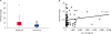
The present study was initiated based on previous literatures, which indicates that a high plasma level of YKL-40 is associated with metastasis and shorter survival in a number of human cancers [81012]. Blood samples were collected from 82 dogs with the following types of cancer: lymphoma (n = 30), mast cell tumor (n = 15), mammary gland tumor (n = 10), melanoma (n = 7), sarcoma (n = 7), adenocarcinoma (n = 6), and others (n = 7; Table 1). Twenty blood samples collected from healthy dogs were used as controls. The YKL-40 levels in blood from healthy dogs and from those with cancer were evaluated using a commercial ELISA kit. The median YKL-40 level was significantly higher (p = 0.0009) in the plasma from 82 dogs with cancer (179.9 pg/mL; range, 39.9–10822.4) than in the plasma from normal healthy dogs (29.99 pg/mL; range, 12.0–263.5). Individual plasma YKL-40 levels in the healthy dogs and in the dogs with cancer are illustrated in Fig. 1.
Table 1
Demography of dogs with cancer in this study

| Types of cancer | Lymphoma | MCT | CMT | Melanoma | Sarcoma | Adenocarcinoma | Others |
|---|---|---|---|---|---|---|---|
| No. of cases | 30 | 15 | 10 | 7 | 7 | 6 | 7 |
![]()
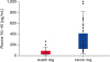 | Fig. 1High plasma levels of YKL-40 are associated with cancer development in dogs. ELISA analysis of 82 dogs with cancer and 20 healthy dogs demonstrates that plasma YKL-40 levels were higher in the dogs with cancer. Two biological replicates were used for each sample, and the data are expressed as the mean values (p = 0.0009, t-test).
|
High levels of plasma YKL-40 predicts a poor prognosis in dogs with cancer
We further investigated the correlation between the YKL-40 plasma level and the prognosis in dogs with cancer. The YKL-40 level was significantly higher in the dogs that later presented with relapse (median, 277.09 pg/mL; p = 0.008) or metastasis (median, 248.58 pg/mL; p = 0.016) than in the dogs that presented no signs of relapse (median, 141.9 pg/mL) or metastasis (median, 155.9 pg/mL; Table 2). The dogs with cancer were subsequently divided into two groups: those with a high YKL-40 level (> 180 pg/mL) and those with a low YKL-40 level (< 180 pg/mL). Relapse and metastasis rates (after treatment) were significantly higher (p = 0.001 and p = 0.04, respectively) in the high YKL-40 group at 67.5% (27/40) and 42.5% (17/40), respectively, than in the low YKL-40 group at 30.1% (13/42), and 21.4% (9/42), respectively (Table 3). These results indicate that the dogs with cancer that presented with high blood levels of YKL-40 had a significantly higher risk of tumor relapse and metastasis when compared with those that had low blood YKL-40 levels.
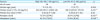
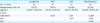
Table 2
Plasma level of YKL-40 and its autoantibody titer in dogs with cancer

Statistical analysis by Student's t-test
YAA, YKL-40 autoantibody.
Significant difference: *p < 0.05; †p < 0.01.
![]()
Table 3
Characterizations of the dogs in the groups of high and low YKL-40 plasma level
YAA, YKL-40 autoantibody.
Statistical analysis by Student t-test (a) and by χ2 test (b); Significant difference: *p < 0.05; †p < 0.01.
![]()
Expression and purification of rcYKL-40 protein
To further investigate the role of YKL-40 in canine tumors, the canine YKL-40 gene was amplified from the macrophage and inserted into the vector pcDNA3.1/V5-HIS-TOPO to create a prcYKL-40 construct. The construct was stably transfected into BALB/3T3 cells to obtain the CL7208 cell, which stably expresses and secretes rcYKL-40. We identified rcYKL-40 in the cytoplasm of CL7208 cells using the anti-His antibody (Fig. 2A), and rcYKL-40 was also detected in the culture medium of the CL7208 cells after proper purification (Fig. 2A and C).
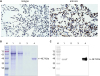 | Fig. 2Expression of rcYKL-40 protein was identified in the CL7208 cells and its culture media. rcYKL-40 was identified in the following manner: (A) in CL7208 cells using immunocytochemistry (probed by anti-His antibody and DAB); in purified supernatants of the CL7208 cell culture media (B) using SDS-PAGE (stained with Coomassie Brilliant Blue); and (C) using Western blot (probed by anti-His antibody).rcYKL-40, recombinant canine YKL-40; CL7208, cells that stably expressed rcYKL-40; DAB, 3,3′-diaminobenzidine; M, prestained protein ladder; Lane 1, concentrated supernatant of CL7208 cell culture; Lane 2, unbind flow-through; Lane 3, wash flow-through; Lane 4, elution flow-through; SDS-PAGE, sodium dodecyl sulfate–polyacrylamide gel electrophoresis.
|
YKL-40 is also an autoantigen in dogs
YKL-40 has been proposed to act as an autoantigen in several human diseases [13]. To examine this aspect of YKL-40 in dogs, the canine sera were employed to probe our rcYKL-40 using a Western blot assay. As shown in Fig. 3, the rcYKL-40 protein was detected by dog sera, which implies that YKL-40 acts as an autoantigen in dogs and can elicit the canine immune system to produce the YAA with different titers.
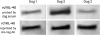 | Fig. 3The sera from three dog (Dog 1, Dog 2, and Dog 3) revealed different titers of autoantibody against rcYKL40 in Western blot analysis. Purified rcYKL-40 (1 µg/well) was separated on 10% SDS-PAGE and transferred to a PVDF membrane, which was probed by three 100× diluted dog sera (Upper lane). The membrane was then stripped and reconfirmed using the anti-His-tag antibody (Lower lane).rcYKL-40, recombinant canine YKL-40; SDS-PAGE, sodium dodecyl sulfate–polyacrylamide gel electrophoresis; PVDF, polyvinylidene difluoride.
|
High titers of YAA in healthy dogs
To determine the correlations between YAA and cancer development in dogs, an indirect ELISA test was performed to detect the YAA titers in the plasma obtained from the dogs. The plasma previously used for the detection of YKL-40 level were reused for the indirect ELISA test (Fig. 4A). YAAs were detected in the blood of both the healthy and the cancer groups; the healthy dogs had significantly higher titers of YAA than the dogs that had cancer (median value, 0.96 vs. 0.38; p = 0.0057). No significant differences in the YAA titers were observed between the dogs with and without relapse or metastasis (p > 0.05; Table 2). Furthermore, no significant differences in the YAA titers were noted between the high YKL-40 and the low YKL-40 groups (p > 0.05; Table 3). However, linear discriminant analysis was used to measure the related linearity to explore the correlation between the plasma level of YKL-40 and its autoantibody. An R2 value of 0.049 (Fig. 4B) was obtained, which suggested a nonproportional relationship between the plasma YKL-40 level and the YAA titer. In addition, no significant difference was found in any of the statistical analyses regarding changes in the YAA titers in the dogs with lymphomas between the cancer and noncancer groups, the relapse and nonrelapse groups, or the high and low YKL-40 groups (Table 4).
 | Fig. 4A low YAA titer in dog plasma is associated with cancer progression. (A) rcYKL-40 (250 ng/well) was coated into each well of a 96-well plate. Diluted (1:50) dog plasma was applied to probe the rcYKL-40. A reading from one healthy dog plasma was defined as 1. The fold change was derived from the ratio of the OD650 absorbance values of the sample to those of the control. Data are shown as the mean ± SD (p < 0.0057, t-test). (B) Linear discriminant analysis revealed nonproportional correlations between YKL-40 and its autoantibody titer in dog plasma.YAA, YKL-40 autoantibody; rcYKL40, recombinant canine YKL-40; SD, standard deviation.
|
Table 4
Plasma YKL-40 levels in dogs with different type of cancer
![]()
DISCUSSION
An elevated YKL-40 plasma level has been correlated with the development and progression of cancer and with poor prognosis in humans [89101112]. The present study provides preliminary evidence of the presence of a similar association between YKL-40 and tumors in dogs. First, the dogs with cancer had significantly higher plasma levels of YKL-40 than the healthy dogs (Fig. 1). The YKL-40 plasma level in the dogs that later presented with relapse or metastasis was higher than in those with no signs of relapse or metastasis (Table 2). Second, the dogs with cancer and with high plasma levels of YKL-40 (> 180 pg/mL) had a significantly higher risk of tumor relapse and metastasis than their low YKL-40 plasma level counterparts. These results suggested that the elevated plasma YKL-40 level might act not only as an indicator of cancer development and malignancy in dogs but also as a useful prognostic marker. To the best of our knowledge, this is the first report on the identification of a possible plasma biomarker, YKL-40, in dogs with cancers.
Although elevated YKL-40 plasma level has been correlated with the development and progression of various human cancer in a lot of studies, each study worked on only single tumor type [89101112]. The difference of plasma levels among different tumor types has not been reported yet. Since the plasma of this study were collected from various types of dog cancer, we further examined the difference of plasma YKL-40 levels among different tumor types. The sources of plasma were grouped into lymphoma (n = 30), mast cell tumor (n = 15), canine mammary tumor (n = 10), and miscellaneous (n = 27). The median values of plasma YKL-40 for each tumor types were listed in Table 4 and we found no significant differences between each other. This indicated the plasma YKL-40 levels may not be a good biomarker to identify specific tumor in dogs. However, the sample size of some tumor types was small in this study. In the future, larger scale study on different types of canine tumor should be done to clarify this issue.
Except for cancer diseases, YKL-40 has been proposed as a pro-inflammatory biomarker in humans and is highly expressed in a variety of human inflammatory diseases of infectious and noninfectious etiology [15161718]. Although we do not know the real role of YKL-40 in canine inflammatory diseases yet, the cases with symptoms of infection were excluded in this study to rule out the influence of inflammation. Dogs with latent infection or inflammation induced by noninfectious diseases may still be involved in this study. However, inflammation has been described as one of the hallmarks of cancer, and it plays an important role in different stages of tumor development and progression [1920]. The accumulation of different types of long-term or chronic inflammation may promote the progression of neoplasm. Persistent high levels of YKL-40 in the blood may be a sign of tumor progression or poor prognosis. Even so, to clarify the role of YKL40 in various canine inflammatory diseases should be important in the future study.
Antibodies are the most favorable choice for plasma screening because of their stability and suitability in sensitive immunoassays. Autoantibodies are produced by the immune system, which is directed against one or more of the individual's own proteins. They were first identified in colon cancer patients more than 40 years ago [21]. Subsequently, tumor-associated autoantibodies against various autoantigens have been detected in a variety of tumors and have been used as diagnostic and prognostic tools [22]. A recent study reported the use of multiplex-tumor-associated autoantibodies to detect lung cancer in the very early stage, months to years prior to diagnosis [23]. Canine cancer research employing autoantibodies has rarely been conducted [24]. YKL-40 has been recognized as an autoantigen, but not yet as a tumor-associated autoantigen [13]. The present study provides evidence of the role of canine YKL-40 as an autoantigen that can elicit YAA in dogs (Fig. 3). Moreover, we found that a low titer of YAA may be related to tumor development (Fig. 4A). However, a significant change in the YAA titer was not observed between good and poor prognostic cases (Tables 2 and 3). In addition, the YAA tilter was not inversely related to the YKL-40 levels in the dogs with cancer (Fig. 4B). Therefore, the YAA titer does not seem to be a suitable cancer marker for dogs thus far. Nevertheless, we believe that the appearance and role of YAA in dogs is an interesting question to explore in the future.
YKL-40 is not known to have a specific receptor, and its biological functions in cancer remain unclear. This study provides preliminary evidence and encourages us to further explore the role of YKL-40 in canine cancer. In the future, large-scale studies to evaluate the changes in blood YKL-40 levels in various tumor types, breeds, and ages and at different stages of tumor progression and treatment will be necessary. In addition, the development of a useful antibody against canine YKL-40 is warranted. Biological functions and signaling elicited by YKL-40 must be investigated in canine cancer cells.
In conclusions, dogs with cancer had higher plasma levels of YKL-40 and lower YAA titers. The plasma YKL-40 levels were significantly higher in the dogs that later presented with relapse and metastasis after treatment. Our results imply that blood YKL-40 levels may have the potential to be developed as a marker of progression and prognosis of canine cancers.
 XML Download