

 PDF
PDF Citation
 Print
Print


INTRODUCTION
Periodontal diseases, such as gingivitis and periodontitis, are inflammatory diseases induced by plaque in the tissues supporting teeth [1]. Periodontal disease is one of the most common oral diseases in small animal medicine [2]. About 80% of dogs and 70% of cats aged 2 years and above have various types of periodontal disease [3]. The importance of dental care in cats is increasing as the awareness of feline dental disease increases. Cats that are severely affected by periodontal disease are reluctant to eat or drink due to severe pain [1]. Studies revealed that small dogs were more vulnerable than large dogs to periodontal disease because of their small teeth, short interdental space, decreased oral chewing, and shorter teeth roots [345].
To manage the dental health of companion animals, owners use many methods, such as brushing teeth, dental hygiene chews, and chewing toys [16]. Especially for dogs, a variety of products for calculus removal, including dental hygiene chews, have been developed, and the effectiveness of plaque removal has been demonstrated by scientific research [7]. On the other hand, there are few products available for calculus removal and dental hygiene in cats. The research on dental hygiene chews for cats has been carried out only to measure the effects of the ingredients on the removal of plaque and calculus. This was studied for human dental hygiene purposes and showed that the shape and size of teeth could affect the accumulation of plaque and calculus [4]. Unlike dogs, cats are obligate carnivores, and both the number and role of their teeth are different from those in dogs. In previous studies, it was shown that physical calculus removal methods for cats such as brushing teeth could prevent periodontitis, and that foods with abrasive and hard textures could effectively prevent plaque and calculus formation if effective contact was made between the food and surface of the teeth [18]. Studies also revealed that dental health could be improved with increased chewing time and increased number of chewing cycles [910111213].
Using the same equation for the average mouth scoring system for all species is not reasonable, because each animal species has different sizes of teeth and mouth size [4]. There are many cat-related products for calculus removal, but no scientific studies have been performed for analyzing the anatomical features of dentition in cats. Therefore, measurement of the size and shape of cats' teeth may be necessary to increase the efficacy of dental care interventions.
The purpose of this study was to investigate the oral structure and mastication mechanism of cats to aid in designing the shapes and materials of dental hygiene chews to effectively remove calculus in cats.
MATERIALS AND METHODS
Ten cats were recruited for the study with informed consent from their owners. There was no restriction on breeds recruited. Age was restricted to one year or older as far as possible to exclude the variance related to age. Cats with dental disease such as teeth loss were also excluded. This research was approved by the Institutional Animal Care and Use Committee of Seoul National University (SNU-190116-2).
Blood analyses and thoracic X-rays were performed for all cats to check their physical health prior to general anesthesia. Cats were premedicated with intravenous (IV) tramadol 2 mg/kg (Huons Tramadol HCl inj. 40 mg/mL; Huons Co., Ltd, Korea) and medetomidine 20 µg/kg (Domitor 0.2 mg/mL; Orion Corporation, Finland); anesthesia was induced with IV alfaxalone 3 mg/kg (Alfaxan; Jurox Pty Ltd, Rutherford, Australia) and maintained with isoflurane in 100% oxygen for dental prophylaxis and dental impression. Yellow stone (Neo Plum Stone; Mutsumi Chemical Industries Co., Ltd, Japan) and alginate (Selection J Alginate Impression; Youdent Co., Ltd, Japan) were used for dental impressions. Teeth-related parameters were directly measured using digital calipers (NA500-200WPS; Blue Bird Inc., Korea) in anesthetized cats. Parameters related to oral size were measured in the dental impressions with digital calipers (Fig. 1). 3rd premolar (PM3) and 4th premolar (PM4) of the maxilla, and PM3, PM4 and 1st molar (M1) of the mandible were measured and defined as parameters related to teeth. The height and width of each tooth was estimated, including the rostral and caudal tip of M1. The interdental spaces between the tips of the teeth (PM3–PM4 in the maxilla, and PM3–PM4, PM4–M1 [rostral tip] and M1 [rostral tip]–M1 [caudal tip] in the mandible) were also measured. As occlusion only occurs in the premolar and molar region, the distance from the tip of PM3 to the caudal tip of M1 in the mandible was measured. Parameters related to oral size were the length between the buccal surface of M1 bilaterally (A) and the length from the mid-point of A to the posterior surface of the incisors (B) in the maxilla and mandible (Fig. 1). The A/B ratio was calculated and the measurements were presented as mean with standard deviation.
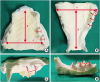 | Fig. 1The parameters measured using digital calipers. (A) maxilla A, B and width of maxillary PM3 and PM4 (*). (B) mandible A, B and width of mandibular PM3, PM4 and M1 (*). (C) the height of maxillary PM3 and PM4 (†), and the interdental space between the tips of each tooth (‡); PM3 and PM4. (D) the height of the mandibular PM3, PM4 and M1 (†), and the interdental space between the tips of each tooth (‡); PM3 and PM4, PM4 and the rostral tip of M1 and the rostral tip and the caudal tip of M1.
|
In order to observe cat's chewing, the cats were offered both types of feeds, generic feeds for over 1 year of age with a size of about 1 cm and feeds for dental care with a size of about 1.5 cm. Videos were recorded during chewing using a digital camera. Lateral and frontal views were recorded to analyze the patterns of chewing motion. The patterns of chewing motion and total chewing time before swallowing were analyzed by reviewing the videos.
Dental hygiene chews considering the anatomical features of the teeth and chewing motion was designed for cats. For image modeling, 3D modeling tool (Rhinoceros; Robert McNeel & Associates, USA) was used.
RESULTS
In this study, 10 cats of 6 different breeds were included: 4 domestic shorthairs, 2 Russian blues, 1 American shorthair, 1 Persian, 1 Turkish Angora, and 1 Devon Rex, all of which were common breeds that accounted for a large proportion of companion cats in Korea. There were no remarkable findings on pre-anesthesia physical examination in any of the cats. Twenty parameters related to teeth and oral size of the 6 breeds were compared and analyzed (Table 1). In the maxilla, A was 36.85 ± 2.94 mm, and B was 34.40 ± 3.34 mm. In the mandible, A was 30.86 ± 2.87 mm, and B was 31.80 ± 2.09 mm. The A/B ratio was 1.07 for the maxilla and 0.93 for the mandible. The largest size of maxilla and mandible in measured cats was 42.0 mm and 34.5 mm. The height of PM3 in the mandible was the lowest (3.40 ± 0.74 mm), and caudal part of M1 in the mandible was the highest (4.50 ± 0.62 mm). The width of M1 in the mandible was the smallest (2.08 ± 0.06 mm), and PM3 in the mandible was the largest (2.17 ± 0.04 mm, Fig. 1).
Table 1
Measurements of each parameter of teeth size and oral size using a digital caliper (mm)

ro., rostral; cd., caudal; A, the length between the buccal surface of M1 bilaterally; B, the length from the mid-point of A to the posterior surface of the incisors; DSH, domestic shorthair; ASH, American shorthair.
![]()
The cats had the following dentition in the maxilla; 3 pairs of incisors, 1 pair of canines, 3 pairs of premolars and 1 pair of molars. The dentition of the mandible was the same as that of the maxilla, except that there were 2 pairs of PMs. All premolars and molars had highly sharpened tips. The lateral views of the dentition showed that the tips of the upper and lower teeth were not in contact with each other during occlusion. Occlusion was observed mainly between PM3 and PM4 in the maxilla and all PMs and M1 in the mandible. The length of the interdental space between PM3 and PM4 in the maxilla was 7.83 ± 0.85 mm, while the length of the interdental space between PM3 and PM4 in the mandible was 6.65 ± 1.31 mm. The length of the interdental space between PM4 and the rostral tip of M1 was 3.80 ± 0.58 mm, and the length of the interdental space between the rostral tip and caudal tip of M1 in the mandible was 5.25 ± 0.48 mm. The longest length of dental occlusion region in measured cats was 18 mm and the mean length was 15.70 ± 1.77 mm (Fig. 2).
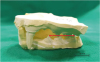 | Fig. 2Representative images of lateral views from a dental impression in cats. The arrow presents the total length of the dental occlusion region; the maxillary PM3 and PM4 and mandibular PM3, PM4 and M1.
|
Analysis of the chewing mechanism revealed that the cats first took the food with the incisors and directly moved it to the premolars and molars for chewing. Usually the cats chewed the food 3 to 7 times before swallowing; this depended on the material and size of the food. The chewing motion was more of a guillotine-like motion rather than a crushing motion. The initial chewing work was the most significant, involving force for breaking the food. The later chewing work provided additional assistance in breaking down the smaller parts of the food. The incisors and canines were rarely used in the chewing exercises. The larger the size of feeds, the greater the chewing activity with molars, but no other chewing pattern was observed except the cutting-pattern like scissor. In this study, the difference in hardness between the 2 types of feeds was not significant and could not be compared.
The surface of the dental hygiene chew could be designed to protrude so that more abrasion occurs between the tooth surface and the crevices of the chews for abrasive on the tooth surface. Based on the above criteria, 4 types of dental hygiene chews for cats could be designed. The first model was designed with an overall size of 18 mm, taking into account only the length of the occlusion region. The second, third and fourth models had the overall size of 45 mm considering the oral size and 18 mm in height considering the dental occlusion region. The difference between the 3 models was the appearance of the surface. The surface shape of the models was designed with consideration of the tooth size (Fig. 3).
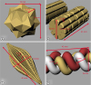 | Fig. 3The expected models of dental hygiene chew according to the results of this study. (A) one mouth size model; The overall size was determined based on the total length of occlusion region, which was the largest measured as 18 mm. This model chew should not be swallowed at once. (B, C) and (D) the width of dental hygiene chew was determined based on the widest length of oral size measured as 45 mm. The height of chew was determined based on the total length of occlusion region measured as 18 mm. The distance between the protrusions was determined as 2 mm considering width of tooth. (D) the protrusion width was determined as 8 mm considering the length of interdental space.
|
DISCUSSION
Cats have a relatively prolonged lifespan following the development of veterinary medicines [13]. Owners' concerns about their health has also increased. Of the various health care concerns, the oral health care of companion animals is very important but difficult to manage. Cats are particularly sensitive to stress, so their oral health care is more difficult to maintain than it is in humans or in dogs. It has been proven that daily brushing of a cat's teeth reduces dental plaque and calculus in their teeth and prevents gingivitis and periodontitis, but practically it is difficult for the owner to maintain the oral care of the cat [1]. In dogs, many reports have already been published showing the effectiveness of dental hygiene chews for oral care [910111214], and various products are actually used in the clinical field. Generally, the friction created between the dental hygiene chew and the tooth surface is known to be an important mechanism to remove plaque and calculus [9]. In cats, it has also been proven that products of abrasive and rough materials are effective in removing plaque on the surface of teeth with the same mechanism [8]. Therefore, this study was conducted to design dental hygiene chews for cats considering the anatomical characteristics of the dentition.
In dogs, there were significant differences in the size and shape of the oral cavity between the dolichocephalic and brachycephalic breeds [7]. Thus, to achieve sufficient physical friction at the tooth surface, the size and shape of the dental hygiene chews must be different according to the skull type of the breeds [7]. According to the results of this study, however, there was not much difference when comparing the teeth sizes and oral sizes of the 6 cat breeds. Even the Persian cats, which represent the brachycephalic breed, did not have much difference in teeth size and oral size when compared to other breeds included. Therefore, unlike dogs, only one type of dental hygiene chew will be sufficient for cats.
Cats are obligate carnivorous animals. For cats, the premolars and the molars play the main role in the chewing activity. They resemble a scissor's blades, and cats use them to cut and tear the food. The appearance of such teeth induces less frequent chewing movements. Even smaller food pieces are not chewed but swallowed easily one at a time in cats. In order to remove plaque and calculus from teeth, dental hygiene chews need to physically scrape the surface of the teeth, so they should be designed to encourage more chewing work from the cats.
The total size of the teeth and oral cavity, as measured with digital calipers, could be effectively used in the design of dental hygiene chew. To encourage chewing activity, the first value to be considered was the overall size of the chews, which was related to the length of the dental occlusion region that was primarily responsible for chewing activity. It has been confirmed that the dental occlusion region consists of the maxillary PM3 and PM4, and the mandibular PM3, PM4, and M1 (Fig. 2). The overall size of the chews should be larger than the dental occlusion region so that minimal chewing activity could be induced without swallowing at once and all involved teeth could be scratched. In this study, the length of the average occlusion region of the cats was 15.7 mm, but the length of the longest occlusion region of the cats was 18 mm. The dental hygiene chews should theoretically be bigger than 15.7 mm. However, the size of the expected dental chew was determined to be 18 mm, the longest length measured, since all cats have to induce chewing activity and stimulate all teeth. The second value to be considered for designing a dental hygiene chew to encourage more chewing activity in cats was the oral size; the length between the buccal surfaces of M1 bilaterally (A) and the length from the mid-point of A to the posterior surface of the incisors (B) in the maxilla and mandible. If the chews were larger than the length of the occlusion region but less than A and B, it could be inserted into the mouth at once, which would not encourage a large amount of chewing activity, depending on the cat. In this study, the A/B ratio in the maxilla and mandible was close to 1, and the mean of the widest width measured in cats was 36.85 mm. However, the largest oral size among the measured cats was 42 mm, which might be difficult to induce more chewing activity for cats with a larger oral size, considering only the average size. Dental hygiene chews should be designed longer than 42mm so that all cats were bigger than their mouth size and could not bite into the mouth.
An important feature of dental hygiene chews is that they cause as much abrasion on the tooth surface as possible. Therefore, it is necessary to increase the area of the friction surface between the dental hygiene chew and tooth surface [18]. The surface of the chew could be designed to protrude so that more abrasion occurs between the tooth surface and the crevices of the chews. The height of protrusion should be no longer than 4 mm according to the heights of the teeth, as it could irritate and damage the cat's own gums if it was more than the tooth height. Considering the width of the tooth, the length between the protrusions could be 2 mm to allow the tooth to enter the crevice space, which would induce each protrusion to make contact between the tooth surface and the dental hygiene chew surface.
Based on the above criteria and the measurement results, 4 types of dental hygiene chews for cats could be designed. The first model was designed to be portably used as a snack, while the overall size encouraged the chewing activity in relation to the length of the dental occlusion region. The overall size was 18 mm and the surface were designed with a star candy-shaped so that the protrusions could physically scraped the tooth surface. In order to induce more chewing activity, the overall size of dental hygiene chew could be considered in relation to oral size. The overall sizes of the second, third, and fourth models were determined by the measured oral size and the dental occlusion region. The width and height of the models were 45 mm and 18 mm. The difference between the 3 models was the appearance of the surface. The second model was a wooden shell-shaped surface, the third model was a toothbrush-shaped surface and the fourth model was a pretzel-shaped surface. It was designed to have crevices on the surface of the models so that the tooth could enter into the crevices and physically scratch the tooth surface. The surface of the fourth model was made with pretzel-shaped protrusions, the length between the protrusions was set to 8 mm considering the length of the interdental space (Fig. 3).
The limitation of this study was the small sample number of cats. As more samples are studied, statistically more accurate results might be obtained. It is also necessary to validate these results in a greater number of cat breeds. Unlike dogs, dolichocephalic and brachycephalic breeds of cat are not clearly distinguishable; hence, it is important to compare a variety of breeds and identify the exact differences in each breed. In order to develop an effective dental hygiene chew, it is also necessary to study the ingredients of chew, for not only the shape of the chew, but the practical aspect of the ingredients should be considered. Studies revealed that materials to create abrasion at the tooth surface during chewing were effective to eliminate dental calculus [18]. In addition, the model should be designed specifically to induce chewing activity and should be made of ingredients that are easy to digest considering the possibility of the device to be swallowed during the chewing process. it is also necessary to increase accessibility of dental hygiene chews with a more favorable taste to cats.
Furthermore, the 4 models of dental hygiene chews made by considering all these factors should be adapted, and further study on their clinical efficacy in cats with dental calculus should be needed.
In conclusions, the oral size and the teeth sizes of cats included in this study were very similar. The overall size of dental hygiene chews could be determined based on the measurements of the oral size. Furthermore, it was recommended that the surface details of the dental hygiene chews be designed taking into consideration anatomical teeth parameters, in order to remove and prevent dental calculus and plaque in cats.
 XML Download