

 PDF
PDF ePub
ePub Citation
 Print
Print


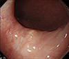
A 64-year-old man was referred to our hospital for a routine medical examination. His past medical history revealed only essential hypertension. He was an ex-smoker and a social drinker. Physical examination was unremarkable. His blood and serum test results were within normal limits. Upper gastrointestinal endoscopy showed a slightly yellow flat nodule mimicking a subepithelial tumor located in the lower esophagus (about 5 cm above the gastroesophageal junction) (Fig. 1). The endoscopic biopsy specimen showed benign acanthosis. The rest of the stomach and duodenum revealed no abnormalities except for mild erythematous gastritis. We performed precutting endoscopic mucosal resection (EMR) with partial circumferential incision after sufficient submucosal injection for diagnostic purposes.
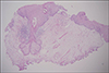
Histopathological examination revealed that some lobules of sebaceous glands were located in the deep portion of lamina propria slightly infiltrated by inflammatory cells. No hair follicles were observed, and there was no evidence of malignancies (Fig. 2).
Sebaceous glands are normally found in association with hair follicles which are derived from the pilosebaceous apparatus. Ectopic sebaceous glands have been found in various tissues of ectodermal origin, such as the palms and soles, nipples, external genitalia, and parotid glands.1 Sebaceous glands are rarely derived from endoderm in the esophagus.23
The presence of sebaceous glands in the esophagus has not yet been completely explained. Several hypotheses have been postulated. Although both embryologic misplacement and the metaplasia of esophageal submucosal mucus glands or squamous epithelium have been proposed, it seems more likely that sebaceous glands in the esophagus originate as a result of acquired metaplasia of squamous epithelium.4
The endoscopic findings may appear as yellowish papules or nodules of various sizes and numbers. A differential diagnosis is needed in the presence of other yellowish subepithelial lesions in the esophagus, such as carcinoid tumor, granular cell tumor, or xanthoma.5 Pathologic confirmation can be more important than endoscopic finding. In most cases, diagnosis is possible with endoscopic biopsy. Occasionally, endoscopic forcep biopsies are insufficient to predict final histology in a single lesion mimicking subepithelial tumor as in our case. A bite-on-bite biopsy or diagnostic EMR is needed because the ectopic sebaceous glands are located in the deep portion of lamina propria.
On the basis of what has been observed so far, ectopic sebaceous glands are entirely benign in nature, and there are no significant changes during 3 years of follow-up.6 There is still a lack of evidence to support the need for further surveillance endoscopies.
 XML Download