

 PDF
PDF ePub
ePub Citation
 Print
Print


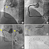
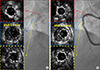
A 68-year old female patient visited our hospital due to effort angina 1 month ago. She had a history of hypertension and diabetes mellitus, and underwent 2 instances of percutaneous coronary intervention (PCI) at right coronary artery (RCA). A 3.5×32 mm Taxus® (Boston Scientific, Natick, MA, USA) was implanted for proximal RCA 14 years ago, and 3.5×28 mm Cypher® (Cordis, Hialeah, FL, USA) was implanted for significant in-stent restenosis (ISR) in RCA 8 years ago (Fig. 1A, 1B). Twelve-lead electrocardiography showed sinus bradycardia, and the level of cardiac biomarkers were within normal range. Transthoracic echocardiography showed normal left systolic function without wall motional abnormality. So, she was diagnosed with unstable angina pectoris and coronary angiog-raphy (CAG) was done and the CAG showed normal left coronary artery system, but significant type-IC ISR in RCA (Fig. 1C). However, fluoroscopic image and StentBoost® enhanced fluoroscopic image revealed complete transection in previous drug-eluting stents' (DES) overlapped site (Fig. 1D, 1E). After wiring into RCA using Runthrough® wire (Terumo, Tokyo, Japan), we examined intravascular ultrasound for further evaluation, and it confirmed complete stent fracture in DES overlapped site (Fig. 2A). As the patient complained effort angina, and minimal lumen area at the stent fracture site were significant (2.8 mm2), we decided to perform PCI for this lesion. After balloon angioplasty using a 3.5×10 mm non-complaint balloon was used to burst pressure (RAIDEN3®, Kaneka, Tokyo, Japan), a 3.5×15 mm Xience Sierra® (Abbott Vascular, Santa Clara, CA, USA) was implanted at the ISR site. However, follow-up CAG showed stent underexpansion, therefore we performed additional balloon angioplasty using a 3.5×10 mm non-complaint balloon. Final CAG showed good distal flow without significant residual stenosis (Fig. 2B).
This case is highly unique in that it was successfully managed using a newer generation DES. Although stent fracture in this case occurred in sirolimus-eluting stent at RCA which was the most common type of DES related to stent fracture and the most common site of fracture, respectively, complete stent fracture at the DES overlapped site is very rare.1 Overlapping coronary stent technique may increase the risk of stent fracture, and most stent fractures were observed near stent overlapping zones.23 It is noteworthy that not all stent fractures are candidates for this treatment.4 However, the patient in the current case complained of recently developed angina and lesion area was significant (reference area in non-left main coronary artery is 4.0 mm2). Furthermore, complete separation in stent segment was known to be related to adverse clinical outcomes with stent thrombosis and ISR than partial stent fracture.3 In case of 1st generation stent fracture needing PCI, newer generation DES seems to be more effective.4 This case emphasizes the importance of stent optimization after PCI, and highlights the usefulness of intravascular imaging and enhanced fluoroscopic imaging to accurately detect stent fracture which was not available using conventional fluoroscopic image.5
 XML Download