

 PDF
PDF ePub
ePub Citation
 Print
Print


INTRODUCTION
Globally, osteoporosis is estimated to affect 200 million women1 and 1 in 3 women over age 50 will experience osteoporotic fractures, as will 1 in 5 men over the age of 50.23 In Korean people aged 50 or older using the Korea National Health and Nutrition Examination Survey (KNHANES) 2008–2011, the prevalences of osteoporosis were 7.0% in men and 40.1% in women.4 Fractures due to osteoporosis are difficult to treat, resulting in tremendous economic losses.5 Also, fractures reduce the quality of life of postmenopausal women by limiting their movement and causing pressure ulcers, cardiopulmonary dysfunction, and venous thrombosis.6 Therefore, prediction and, if possible, prevention, of osteoporosis are of considerable importance.
The inflammatory response protects the damaged area and maintains homeostasis.7 However, if this reaction persists, long-term secretion of inflammatory mediators can induce chronic inflammatory diseases.8910 Chronic inflammation is also present in vascular diseases, and is used as to predict their development.1112 Additionally, osteogenic factors are secreted during chronic inflammation to induce osteoporosis.1314
As with inflammatory measures, the white blood cell (WBC) count, C-reactive protein (CRP) level, and erythrocyte sedimentation rate (ESR) are useful in clinical practice, but have low diagnostic specificity.15 Recently, the neutrophil to lymphocyte ratio (NLR) and platelet to lymphocyte ratio (PLR) have been known as new markers of the systemic inflammatory response.1617 Specifically, the NLR and PLRs are predictive of an inflammatory response in the vascular system17 and more accurate in this regard than the level of CRP, a predictor of myocardial infarction and mortality.18 Also, some researchers reported that the NLR and PLR are associated with a decreased BMD and osteoporosis.1920 However, the relationship between the NLR and PLR and osteoporosis have yielded inconsistent results. All this said, to the best of my knowledge, there has been no study of the association between the NLR and PLR and osteoporosis in the Korean population. Thus, we investigated the relationships among the NLR, PLR, and lumbar and femoral neck BMD in postmenopausal Korean women.
MATERIALS AND METHODS
1. Participants
This study involved postmenopausal women admitted to an orthopaedic hospital in Korea from January 2016 to August 2018. During this period, 498 postmenopausal patients were able to perform the activities of daily living and to communicate. Of this group, 407 were enrolled after we excluded 91 with fractures or a history of treatment for osteoporosis that could affect the test results. Patients who were admitted to the hospital during the study period (February 2018 to August 2018) were interviewed by the researchers and patients who had been discharged from the hospital during the study period were interviewed when they visited for outpatient treatment. This study was conducted in accordance with the Declaration of Helsinki, and the protocol was approved by the Institutional Review Board of Chosun University Hospital (no: 2018-04-015) and informed consent was obtained from all patients.
2. Measures
Experienced investigators reviewed the subjects' medical records. When the information therein was insufficient, a telephone survey was conducted to obtain information on current smoking, monthly drinking, exercise, daily coffee consumption, hypertension, diabetes, use of analgesics, use of antibiotics, use of nutritional supplements, use of steroids, and history of surgery. The current smoking status was assessed by self-report; subjects were classified as current smokers based on their current smoking habits. Monthly drinking was assessed by exploring drinking behaviour during the month prior to interview. Exercise was coded yes when a subject walked for >30 min more than five times per week. Hypertension and diabetes was coded yes when a subject was treating for hypertension and diabetes.
Venous blood samples were collected at the first visit to the hospital. Differential WBC counts were performed using the automated ADIVA® 120 Hematology System (Bayer Corporation, Tarrytown, NY, USA). The NLR was calculated by dividing the number of neutrophils by the number of lymphocytes, and the PLR was calculated by dividing the platelet count by the number of lymphocytes.
The lumbar spine and femoral neck BMD were measured by dual energy x-ray absorptiometry using a DEXXUM-T instrument (Osteosys, Inc., Seoul, Republic of Korea). The lumbar spine BMD was calculated as the mean of the values of the L1–L4 spine segments. Daily phantom scans were performed each morning for quality control purposes, and all BMD scans were conducted by trained examiners using standardized procedures according to the manufacturer's recommended protocols. All BMD scans were reviewed by an experienced investigator to ensure that the region of interest was defined appropriately.
3. Statistical analysis
Statistical analysis was performed using SPSS version 22.0 (SPSS Inc.; Chicago, IL) for Windows. Data is expressed as frequencies and percentages or means±standard deviations. The associations of NLR and PLR quartiles with the subjects' characteristics were evaluated. Analysis of covariance (ANCOVA) was performed to identify significant differences in the lumbar spine and femoral neck BMD according to NLR and PLR quartile. Model 1 was not adjusted, and Model 2 was adjusted for age, height, weight, current smoking, monthly drinking, exercise, and daily coffee consumption. Model 3 was adjusted for the Model-2 variables plus hypertension, diabetes, use of analgesics, use of antibiotics, use of nutritional supplements, use of steroids, and history of surgery. A value of p<0.05 was considered indicative of statistical significance.
RESULTS
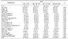
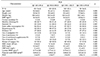
Table 1 shows the subjects' characteristics according to NLR quartile. Age, daily coffee consumption, WBC count, CRP level, lumbar BMD, and the PLR differed significantly according to NLR quartile. Table 2 shows the characteristics of the subjects according to NLR quartile. The ESR and NLR differed significantly according to PLR quartile.
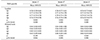
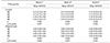
Table 3 lists the mean (95% confidence interval [CI]) lumbar and femoral neck BMD according to NLR quartile. The lumbar BMD decreased significantly as the NLR quartile increased after adjusting for other covariates (Q1, 0.79 [0.77–0.81]; Q2, 0.77 [0.75–0.79]; Q3, 0.76 [0.74–0.78]; Q4, 0.74 [0.72–0.77]; p=0.040, p for trend=0.005). However, the femoral neck BMD was not associated with the NLR quartile. Table 4 lists the mean (95% CI) lumbar and femoral neck BMD according to PLR quartile. Neither the lumbar BMD nor the femoral neck BMD was associated with the PLR quartile.
DISCUSSION
We investigated the relationship between BMD and the NLR and PLR in Korean postmenopausal women. After adjusting for confounding variables, the NLR quartile was negatively associated with the mean lumbar BMD, but the PLR was not associated with BMD.
In the present study, the lumbar spine BMD decreased significantly as the NLR increased. In a study of 1,635 patients aged 65 years or older with or without osteopenia or osteoporosis, a multivariate logistic analysis showed that osteoporosis increased as the NLR increased.19 Among 233 postmenopausal women without diabetes, a multivariate logistic analysis showed that osteoporosis increased as the NLR increased.21 In a study with 438 female patients who visited a hospital, both the lumbar spine and the femoral neck BMD decreased as the NLR increased,22 which is in agreement with our findings.
In our study, there was no significant relationship between the PLR and BMD. Few previous studies have assessed the relationship between BMD and the PLR. One study investigated the associations among the PLR, NLR, and BMD in 211 postmenopausal women who visited a hospital; the PLR was negatively correlated with BMD, whereas the NLR was not.20 However, only a simple correlation analysis, not a multivariate analysis, was performed. In a study involving 4,120 patients, the level of vitamin D, which is associated with BMD, significantly decreased as the PLR increased, but the relationship between the NLR and vitamin D was not significant.23 However, because that study did not analyze the association of the NLR and PMR with BMD, the results cannot be directly compared with our findings.
The mechanisms by which chronic inflammatory factors, including the NLR and PLR, affect BMD are known. Inflammatory cytokines act on mesenchymal stem cells and osteoclast precursors to increase osteoclast-mediated bone resorption.13 These cytokines bind to stromal cells, increasing the production of receptor activator of nuclear factor-kappa B (NF-ĸB) ligand (RANKL) and macrophagecolony stimulating factor and decreasing that of osteoprotegerin (OPG); together, these effects increase the activity of osteoclasts.24 Furthermore, the reduced estrogen level in postmenopausal women induces secretion by the T cells of a variety of inflammatory cytokines, which promotes the production of factors that activate osteoclasts, leading to the development of osteoporosis.25
We found that the NLR, but not the PLR, was related to BMD. The predictive utility of the NLR and PLR differs according to the chronic inflammatory condition in question. In patients with end stage renal disease,26 the PLR has greater predictive power than the NLR. In contrast, the NLR has greater power to predict the prognosis of patients with gastric cancer27 and is a superior indicator of mortality in patients with breast cancer.28 The different predictive utilities of the PLR and NLR are likely caused by the different actions of platelets and neutrophils. Neutrophils secrete the receptor activator of NF-ĸB (RANK), RANKL, and OPG,29 and their interaction with osteoclasts activates bone resorption;30 therefore, the NLR is more strongly associated with BMD than is the PLR.
Several studies have evaluated the association of the CRP level with BMD. In a US study of the relationship between the CRP level and BMD in 2,087 65-year-old women, a simple correlation analysis indicated a significant correlation between the CRP level and BMD (p<0.001), but this relationship disappeared upon multiple regression analysis.14 In Sweden, a cohort study of 1,044 women aged 75 years or older showed no association between the CRP level and BMD.31 However, in a study using the US 1999–2004 National Health and Nutrition Examination Survey data (n=10,475), BMD decreased significantly as the high-sensitivity CRP (hsCRP) level increased in both males and females.32 In our further analysis, the WBC count, ESR, and CRP level were not significantly associated with the lumbar spine BMD or the femoral neck BMD (data are not shown).
This study had the following limitations. First, its cross-sectional design precluded determination of the causality of the relationships identified. Second, we did not analyze other factors that could affect BMD, such as the levels of vitamin D and parathyroid hormones. However, for the first time, there was a meaningful investigation of the relationship between NLR, PLR and BMD in Korean postmenopausal women.
In conclusion, after adjusting for confounding variables, the NLR quartile was negatively associated with the mean lumbar BMD in Korean postmenopausal women. However, the PLR was not associated with BMD.
 XML Download