

 PDF
PDF ePub
ePub Citation
 Print
Print


INTRODUCTION
Cardiac fibrosis is a typical phenomenon that occurs during cardiac muscle remodeling due to various heart diseases such as myocardial infarction (MI) and cardiac hypertrophy. Many experimental mouse models, such as permanent or temporary coronary occlusion, are commonly used in the field of cardiovascular research to increase cardiac fibrosis in the myocardium.12 In these models, myocardial remodeling occurs as MI progresses. This is accompanied by the formation of fibrotic tissue, which plays a decisive role in determining myocardial systolic and diastolic function.3 Therefore, the detection and quantification of myocardial fibrotic tissue after post-MI is an important factor in myocardial remodeling analysis.
In clinical practice, MI is undergoing various treatments such as cardiovascular surgery, stent implantation, and thrombolysis, and there is a lot of interest and research into secondary prevention through many medications.4 After reperfusion therapy for MI, β blockers, angiotensin converting enzyme inhibitors and angiotensin receptor blockers (ARBs) are used to prevent cardiovascular ischemia and to maintain cardiac function.567
Dukarb® (Boryung Pharmaceutical Co., Ltd., Seoul, Republic of Korea) is a fixed-dose combination of fimasartan (ARB) and amlodipine (calcium channel blocker). Recent clinical and experimental research has shown that ARBs have a cardioprotective effect that exceed their ability to lower blood pressure (BP), such as anti-vascular agents, and target organ protection.8 In addition, amlodipine is a long-lasting, dihydropyridine CCB that is now widely used for cardiovascular disease in lowering BP. Recently, amlodipine has been effectively used in combination with other classes of antihypertensive agents to reduce BP and cardiovascular risk.9
So far, little data is available about the effects of fimasartan/amlodipine fixed-dosed combination (F/A) on the cardioprotective effects. Therefore, the aim of this study was to evaluate the effects of F/A on left ventricular (LV) systolic function and infarct size in a rat MI model.
MATERIALS AND METHODS
1. Study animals
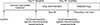
Sprague-Dawley male rats (Samtako, Seoul, Republic of Korea), weighing 250–260 g, aged 8 weeks, were used in all experiments. We induced MI in 20 rats and they were divided into two groups [MI group (n=10) vs. MI+F/A 10 mg/kg group (n=10)] (Fig. 1). The Ethics Committee of the Chonnam National University Medical School and the Ethics Committee of the Chonnam National University Hospital (CHU IACUC-H-2017-23) approved animal experiment protocol.
2. MI model
MI was induced with a slightly modified Fliss method.10 Sprague-Dawley rats were anesthetized with femoral intramuscular injection with a ketamine (50 mg/kg) and xylazine (5 mg/kg). Anesthetized rats were fixed to the operating table with tape. In addition, the respiration was continuously maintained through the ventilator (Model 683, Harvard apparatus, USA). To induce MI, the middle left anterior descending coronary artery was ligated with 5-0 silk to the myocardial region. Finally, the heart was originally positioned in the thoracic cavity, and closed the skin with 1-0 silk. A heating pad (temperature at 37±0.5℃) was used to maintain the body temperature and to increase the survival rate of the rats.
3. Drug administration
F/A (30/5 mg) was pulverized in a tablet grinder and resolved in 10 ml of water. In addition, the vortex (Vortex-Genie 2, Scientific Industries) was used for uniformity of drug before administration of F/A. The body weight was measured daily before the administration of the drug and was administered proportionally to the body weight (10 mg/kg). The F/A was administered by oral injection using zonde needles (1.2×80 mm, 18 G) for 28 days between day-7 and day-35 after the induction of MI (Fig. 1).
4. Echocardiography
A conventional echocardiography was performed according to the main laws of the American Society of Echocardiography.11 Echocardiography was checked in supine position after shaving the chest hair of the subjects. We acquired short-axis images of the LV by transthoracic echocardiography, M-mode and two-dimensional echocardiography images were obtained from the papillary muscle level using echocardiography machine (15-MHz linear array transducer, iE33 system, Philips medical systems).12 Echocardiography was performed at baseline, at day-7, and at day-35 (Fig. 1).
5. Measurement of infarct size
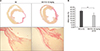
After opening the thoracic cavity, cardiac arrest was induced by administering potassium perchlorate (KClO4) to the thoracic aorta. After heart tissue extraction, saline was used to remove blood from the LV. Cardiac tissue was fixed in 4% paraformaldehyde solution for 3 days, and fixed in paraffin. The tissue fixed to the paraffin was cut to a thickness of 4 µm. Picrosirius red staining was performed to confirm the fibrotic tissue and scar formation in the heart. The infarct size was measured using image analysis program for Image J.13
6. BP measurements
Systolic BP was measured with the tail-cuff method (Visitech Systems, BP 2000).14 Rats were trained to be able to measure BP more than three times per week before drug administration. Systolic BP was measured when the rats were awake before they were scarified at day-35.
7. Statistical analysis
We used Statistical Package for the Social Sciences (SPSS) 22.0 for Microsoft Windows (SPSS, Inc., Chicago, IL, USA) for all statistical analyses. All numerical variables were presented as mean value±standard deviation (SD) and were compared by independent samples t-test. A p value <0.05 was considered statistically significant.
RESULTS
1. Mortality rate
The mortality rate after MI induction was 40% overall, and the mortality rate during the follow-up period was about 30%.
2. Evaluation of LV function by echocardiography
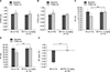
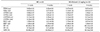
LVEF was measured in a total of twenty rats. A LV functional evaluation was assessed at day-7 and day-35 follow-ups after the induction of MI (Table 1). LV internal diameter at diastole and LV internal diameter at systole were similar between the MI group and the MI+F/A 10 mg/kg group (Fig. 2A, B). Fractional shortening was significantly greater (16.18±1.99% vs. 12.30±2.17%, p<0.001) and LVEF (38.10±3.92% vs. 29.86±4.56%, p<0.001) was significantly higher in the MI+F/A 10 mg/kg group than the MI group (Fig. 2C, D). The delta EF was obtained by subtracting the day-7 measurement from the day-35 measurement. Delta EF was significantly higher in the MI+F/A 10 mg/kg group than the MI group (0.14±2.66% vs. −8.53±2.49%, p<0.001) (Fig. 2E).
3. BP measurements
Systolic BP was not significantly different between the MI group and the MI+F/A 10 mg/kg group at day-7 after the induction of MI (110.11±14.82 mmHg vs. 111.00±13.35 mmHg, p=0.87). There was no significant difference in pulse rate between the MI group and the MI+F/A 10 mg/kg group (381.07±50.54 BPM vs. 386.17±43.21 BPM). However, at the day-35 follow-up, systolic BP was significantly decreased in the MI+F/A 10 mg/kg group compared to the MI group (103.23+13.35 mmHg vs. 123.43+14.82 mmHg, p<0.01).
4. Measurement of scar tissue and infarct size
High color contrast is an important factor in the measurement of fibrotic tissue. Scar formation of cardiac muscle after MI was identified as pricosirius red staining (Fig. 3A) which is marked as red in the fibrotic tissue within the scar in the heart muscle, and is marked as yellow in the non-infarcted tissue. The infarct size was quantified using the Image J program. The MI+F/A 10 mg/kg group had a significantly smaller infarct size than the MI group (26.84±5.31% vs. 36.79±3.10%, p<0.01) (Fig. 3B).
DISCUSSION
The present study was conducted to evaluate the effects of F/A on LV systolic function and cardiac fibrosis in a rat MI model. F/A fixed-dose combination treatment after the induction of MI significantly improved LV systolic function and significantly reduced infart size.
After MI development, the heart undergoes a series of pathologic remodeling processes, resulting in heart failure and cardiac death.15 After MI, myocardial reconstruction can have a significant impact on the function of LV and prognosis for survival. The expansion of the infarct is followed by a long-term growth phase of fibroblast proliferation and collagen deposition. Also, the longer the period, the more collagen deposition is accumulated and the infarct area becomes thin and elongate.16
The ventricular enlargement process for early MI can be influenced by three interdependent factors : infarct size, infarction healing, and ventricular wall stress.17 Angiotensin II is signaled through the angioensin II type 1 receptor (AT1) and angiotensin II type 2 receptor (AT2).18 Recent studies have indicated that AT2 receptor stimulation interferes with the deleterious effects of AT1 signaling in fibrotic diseases.19 Activation of this compensation system reduces inflammation and collagen deposition.20
Fimasartan (BR-A-657-K, KANARB®) is a new ARB developed for the first time in Korea. It is a selective AT1 receptor blocker and has shown rapid and potent antihypertensive effects in clinical trials.21222324 Han et al.8 reported that fimasartan preconditioning has the potential to suppress myocardial ischemia/reperfusion injury and these beneficial effects could prevent the mitochondrial dysfunction and apoptosis accompanied by ischemia/reperfusion injury. Lim et al.25 reported that fimasartan attenuates cardiac remodeling and dysfunction in rats after MI and may prevent the progression to heart failure after MI and myocardial fibrosis and inflammation were downregulated by fimasartan.
Amlodipine is a dihydropyridine CCB that is widely used for the treatment of hypertension. Clinical and experimental studies have shown that amlodipine has some BP-independent effects, including reducing risk of adverse cardiovascular events in patients with coronary heart disease without hypertension, attenuating endothelial dysfunction, and alleviating ischemia-reperfusion-induced heart injury.262728
The present study demonstated that F/A fixed-dose combination therapy improved LV systolic function and reduced infarct size effectively after the induction of MI. The cardioprotective effects of F/A seems to result from the additive beneficial effects of fimasartan and amlodipine for cardiac fibrosis and collagen deposition, vascular inflammation, and endothelial function. The present study suggests that F/A treatment can be used as an effective treatment option for preventing LV remodeling and improving LV function in patients with MI.
There are several limitations to be mentioned. First, the study group had to be divided into four groups including a fimasartan only group and amlodipine only group for the evaluation of the synergistic effects of fimasartan and amlodipine. Second, we only focused on the morphological changes of the heart. Furthermore, the molecular part of the morphological change was not evaluated. Third, we observed the effects of F/A on LV systolic function and cardiac fibrosis for only 1-month after the induction of MI. Therefore, further long-term study is needed.
In conclusion, oral administration of F/A 10 mg/kg could improve LV systolic function, lowered blood pressure and reduced infarcted tissue area in rat MI model suggesting its clinical utility as an effective treatment option in patients with MI. Further investigations regarding this drug have to be conducted and its efficacy remains to be proven in larger animal models.
 XML Download