

 PDF
PDF Citation
 Print
Print


INTRODUCTION
A subdural hemorrhage (SDH) is a common neurological disease in elderly patients [1]. Most SDHs resolve with or without minimal sequelae. In participants over than 90 years, SDHs are associated with a poor outcome [2]. In addition, a poor SDH outcome is related to initial neurological status and treatment options [23]. The typical clinical and radiological course of a patient with an SDH may be urgent in the acute stage, followed by postoperative recovery; it rarely progresses to the chronic stage.
In the present case report, we describe a patient with an SDH who was treated surgically and resolved radiologically but who experienced delayed extensive leukomalacia and showed radiologically worse findings in the chronic stage.
CASE REPORT
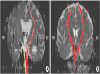
A 62-year-old man presented with slowly progressive dysarthria and left hemiplegia for 1 week without definite acute trauma. He was an alcoholic without any medical history such as high blood pressure or diabetes mellitus; however, his initial brain computed tomography (CT) scan showed an SDH (Fig. 1). Subsequently, the patient underwent burr hole trephination for hematoma removal. Brain CT scans performed on postoperative days 3 and 7 revealed that the SDH had regressed (Fig. 1), and the patient was referred to the rehabilitation department. On day 14, brain magnetic resonance imaging (MRI) showed additional SDH regression (Fig. 2A). At 12 weeks after the onset, the patient's weakness and functional status did not improve. The follow-up MRI revealed extensive leukomalacia, especially in the white matter (Fig. 2B). The diffusion tensor imaging (DTI) for corticospinal tracts (CSTs) revealed severe injury of CST integrity (Table 1, Fig. 3). DTI was performed using a 3.0-T MRI (MAGNETOM® Verio; Siemens, Erlangen, Germany) equipped with a six-channel head coil [4]. The data were acquired in the form of single-shot spin-echo echo-planar images, with axial slices covering the whole brain across 76 interleaved slices of 2.0 mm thickness (no gap); repetition time (TR)/echo time (TE) = 14,300/84 ms; field of view = 224 × 224 mm2; matrix 224 × 224; voxel size 1 × 1 × 2 mm3; number of excitations = 1. Diffusion sensitizing gradients were applied in 64 noncollinear directions with a b-value of 1,000 ms/mm2. The b=0 images were scanned before the acquisition of the diffusion-weighted images, with 65 volumes in total [5]. Fiber tracking was based on the fiber assignment continuous tracking algorithm and multiple regions of interest (ROIs) approach using DTI-Studio. The selection of ROI, termination criteria, and the acquisition of normalized fractional anisotropy (FA) values were proceeded by the same way of previous research [5]. He did not re-gain muscle strength and functional independence, despite 3 months of inpatient rehabilitation. The Mini-Mental Status Examination score was 18 out of 30, and his modified Barthel Index score was estimated to be 45 on a 100-point scale; thus, the patient was returned to the nursing care unit.
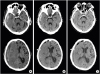 | Fig. 1Brain computed tomography scans over time. (A) Day of onset. (B) Postoperative evaluation on day 4 after onset. (C) Day 7 after onset.
|
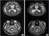 | Fig. 2Brain magnetic resonance images over time. (A) Two weeks after onset. (B) Twelve weeks after onset.
|
Table 1
FN, FA (mid-pons), and FA (pontomedullary junction) values of the CST in case

| Variables | Normalized FN | FA (mid-pons) | FA (pontomedullary junction) |
|---|---|---|---|
| Normalized values | 0.017 | 0.390 | 0.460 |
Values are the normalized values (affected/non-affected).
CST, corticospinal tract; FA, fractional anisotropy; FN, fiber number.
![]()
DISCUSSION
Two clinical aspects are emphasized in the present case report: the occurrence of extensive injury in the chronic stage with extensive white matter injury and relatively preserved gray matter. The patient did not have a history of brain injury or medical comorbidity. He was an alcoholic, his functional status was independent, and organic brain damage was not evident. To date, late extensive white matter injury has not been reported, and, to the best of our knowledge, this is the first description of late extensive white matter injury caused by an SDH. We traced the pathophysiology for delayed extensive white matter injury, and severe motor weakness, the CST injury assessed by DTI in chronic phase would be the explanation for his clinical progress.
An SDH does not always have a benign course, especially in elderly persons older than 70, 85, or 90 years [267]. Typical surgical evaluations such as burr holes or craniotomy induce good clinical outcomes [3]. The patient in this case was not very old and received surgical treatment. At 2 weeks after surgery, imaging studies showed resolution of the SDH. MRI at 12 weeks after the onset had worsened than MRI at 2 weeks after the onset; therefore, delayed brain injury may explain the poorer outcome of SDHs. Physicians should be aware of a possible delayed effect and not disregard neurological changes in the chronic stage of an SDH.
The white matter injury occurred by various mechanisms in SDH. Hematoma itself compressed the CST and induced distortion of the CST. Thus, the reversibility of the FA by DTI was related to the distortion of the pyramidal tract and vasogenic edema due to the expansion of the hematoma in SDH [8]. In addition, secondary insult after SDH was associated with significant brain swelling and stimulated a refractory rise intracranial pressure (ICP). In traumatic SDH complicated by secondary insult, brain swelling is exacerbated by surgical evacuation [9]. An important contributor to the neurological injury associated with SDH is the ischemic damage which is caused by raised ICP producing impaired cerebral perfusion. The removal of the SDH results in the immediate reversal of global ischemia accompanied by an abrupt reduction in the mass lesion and an ensuing reperfusion injury [10].
A previous study found that, in patients with a corona radiata injury, the integrity of the CST, as assessed by DTI obtained during the early stage, appears to be helpful in predicting motor outcomes on the affected side [11]. Most previous reports have shown that damage to the CST is useful for predicting poor outcome of motor function in patients with stroke [111213]. A recent study demonstrated that the CST integrity assessed by DTI, would reflect the hand function up to 12 months after stroke [5]. A previous case report showed CST integrity assessed by DTI may be a useful add-on study for patients with diffuse axonal injury after traumatic brain injury [14]. In this case, the CST integrity assessed by DTI also reflect the motor function and disability in chronic phase in patient with SDH. The CST integrity assessed by DTI would be useful for patients with poor recovery in acute or chronic stage of SDH, despite usual benign course of SDH.
In conclusion, although an SDH is a common neurological disorder and usually has a benign course, delayed white matter injury can occur. Physicians should be aware of the possible delayed effects of SDHs in patients who do not recover or recover slowly. These concerns play an important role in predicting prognosis and establishing a long-term plan for patients and caregivers.
 XML Download