

 PDF
PDF ePub
ePub Citation
 Print
Print


Introduction
The study of the maxillary sinus is crucial for dental surgeons due to its proximity to the teeth and nearby important anatomical structures. The proximity of the roots of innervation of both can lead to pathological alterations in the maxillary sinus, causing symptoms in teeth. Furthermore, inflammatory processes of odontogenic origin may affect the integrity of the maxillary sinus floor.1
Noteworthy abnormalities of the maxillary sinus include the presence of localized or diffuse sinus mucosal thickening, mucosal retention pseudocysts, polyps, nonspecific opacifications (related to acute or chronic sinus inflammatory processes), antroliths, and periostitis due to osteolytic lesions inflammatory lesions.2,3,4,5
Imaging exams are essential for the evaluation and diagnosis of the maxillary sinus, and panoramic radiography (PR) and cone-beam computed tomography (CBCT) should be highlighted as diagnostic modalities. PR is the 2-dimensional radiographic examination most commonly indicated for evaluations of the maxillofacial complex because it is easy to perform, requires a low dose of radiation, and has a low cost. However, its limitations, such as overlapping of structures, uneven magnification, and distortion of the image, can lead to an unfaithful representation of the anatomy and possible pathological alterations, potentially leading to incorrect diagnoses.6 In addition, clinically important areas, such as the anterior wall of the maxillary sinus, may be located outside the cut-off plane of the device, becoming distorted or unseen.5
The advantages of 3-dimensional CBCT over conventional PR include the image quality of high-contrast structures (maxillofacial bone tissues) without geometric distortion or any overlap of the surrounding anatomical structures.6 Therefore, CBCT has become the modality of choice for diagnosis in several situations in dentistry. However, it is important to emphasize that its use instead of PR must be justified considering its higher radiation dose and cost.7
Previous studies have compared the diagnostic ability of PR and CBCT for assessment of the maxillary sinus by clinical dental surgeons and specialists in dental radiology,3 surgery,8 and periodontics.5 According to Dau et al.,8 professional ability directly influenced the performance of imaging tests for the diagnosis of abnormalities in the maxillary sinus. However, the literature is limited in terms of studies comparing the diagnostic ability of these exams when evaluated by dental students.
Given the importance of imaging exams for evaluation of the maxillary sinus and correct diagnoses by dental surgeons, the aim of the present study was to compare dental students' diagnostic ability for sinus abnormalities in PR and CBCT exams.
Materials and Methods
The present study was performed after approval by the Ethics Committee in Research of the Federal University of Juiz de Fora, Juiz de Fora, Minas Gerais, Brazil (register number 2.085.036/2017), and it complied with the recommendations of the National Health Council Ministry of Health of Brazil for research in human subjects.
Sample selection
PR and CBCT images of 150 patients, regardless of sex, race, and age, were selected from the digital image database of a dental radiology clinic of a public university. To be included in the sample, the images were required to have been obtained at the same time, with a clinical indication independent of the present study, and the CBCT images were required to have a field of view (FOV) of 8 cm×13 cm comprising the entire maxilla (teeth and complete extension of the maxillary sinuses). Images of patients with a bone graft in the posterior maxilla and/or the presence of pathological alterations suggestive of cysts, tumors, or fibro-osseous lesions in the maxillary sinus, as well as maxillary fractures, were excluded. After the application of these criteria, 10 patients were excluded: 6 due to incomplete visualization of the maxillary sinuses in the FOV of CBCT and 4 due to the presence of an image suggestive of periosteal osteoma in the maxillary sinuses, resulting in a final sample of 140 patients (280 maxillary sinuses).
Acquisition of images
All CBCT images from the database were obtained with a i-Cat® Next Generation device (Imaging Sciences International, Hatfield, PA, USA) with the patient in maximal habitual intercuspation and positioned according to the light indications of the tomograph (median sagittal plane perpendicular to the ground and ala-tragus line parallel to the ground), with the following protocol of acquisition: 37.07 mAs, 120 kVp, FOV of 8 cm×13 cm, voxel size of 0.25 mm, and scanning time of 26 s with 360° rotation. PR was obtained using an Orthopantomograph® OP100 (Instrumentarium Dental, Tuusula, Finland) with the patient with a semi-open mouth and positioned according to the light indications of the device, with kVp and mA settings chosen according to the patient's biotype.
Classification of sinus abnormalities
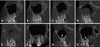
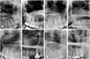
The 280 maxillary sinuses were evaluated according to the classification proposed by Nunes et al. (Figs. 1 and 2);3 1) normal: radiolucent, intact cortical bone, mucosal thickness <3 mm, 2) mucosal thickening: area without cortical bone and with soft tissue density, thickness >3 mm, parallel to the sinus bone wall, 3) sinus polyp: area with soft tissue density forming an extension (fold) adjacent to thickened maxillary sinus mucosa, 4) antral pseudocyst: area with soft tissue density and no cortical bone, dome-shaped, intact sinus floor, 5) nonspecific opacification: soft tissue density, partial or total homogeneous maxillary sinus opacification, 6) periostitis: thick and homogeneous opaque area, laminated, adjacent to the cortical bone of the maxillary sinus floor, above a radiolucent area associated with the tooth apex, 7) antrolith (antral calcification): well-defined radiopaque area, typical characteristics of calcification, and intact maxillary sinus cortical bone or cortical bone within the maxillary sinus. The category of antrolith associated with mucosal thickening was also added.
The maxillary sinuses presented in Figures 1 and 2 represent imaging results of the same structures in CBCT and PR. Each of the categories listed above was classified for each maxillary sinus according to the following 5-point scale: 1: definitely absent, 2: probably absent, 3: uncertain, 4: probably present, 5: definitely present.
Image evaluation
Elaboration of the reference standard
The CBCT images were first evaluated individually by 2 oral radiologists, with more than 8 years of experience, who classified the maxillary sinuses according to the categorization described above. Discordant cases were reassessed together to reach consensus. The final evaluation was used as a reference standard for comparisons with the evaluations performed by students of both the CBCT and PR images.
Evaluation of images by dental students
A pilot study involving application of the proposed methodology in a small sample (18) students was conducted to determine the number of students eligible to participate in the study. Six students did not feel comfortable about the evaluations and refused to participate in the final study, and 10 students were not eligible. Thus, 2 students were included as evaluators of the images, and this number was considered significant for the statistical analysis.
All images were evaluated individually by 2 undergraduate dental students, who had already completed the relevant coursework (oral radiology I and II). They were previously instructed by an oral radiologist about standardization of the evaluations to clarify the purpose of the study, the importance of performing the analyses in a standardized environment, the classification to be used, and how to complete the evaluation spreadsheet. The students then completed to a pilot test (implementation of the methodology proposed in 20% of the sample; n=56 maxillary sinuses) to calculate the intraexaminer and interexaminer reliability through the kappa test, in order to ensure that they could be included as evaluators in the present study. The sample used for the pilot test was not part of the sample used for the evaluation in this research.
All evaluations were performed on a 21.5-inch LCD monitor with high-definition resolution (1920×1080), Dell S2240L (Dell Computadores do Brasil Ltda., Eldorado do Sul, Rio Grande do Sul, Brazil), located in a room with the ambient light off and standardized observation conditions. The PR and CBCT images were randomized and then assessed individually at different times. First, all PR images were evaluated in Windows Photo Viewer version 10.0.17763.1 (Microsoft Corporation, Redmond, WA, USA). It is known that radiologists need to check the radiolucency or opacity based on the surrounding structures and the same structure on the other side; therefore, the whole PR image was assessed, not only the maxillary sinus region as shown in Figure 2. Fifteen days after evaluation of the PR images, the CBCT images were evaluated on a dynamic basis in the XoranCat® software, version 3.0.34 (Xoran Technologies, Ann Arbor, MI), always analyzing the coronal, sagittal, and axial reconstructions. The evaluators were instructed to evaluate a maximum of 20 images per day to avoid visual fatigue and consequent impairment of the evaluations. The following image enhancement tools were used when needed: zoom, brightness, and contrast. Thirty days after performing all the measurements, 20% of the sample was re-evaluated in order to determine the intraobserver and interobserver reliability of the examiners.
Statistical analysis
In order to verify the agreement of students' evaluations of the PR and CBCT images in relation to the reference as follows: values <0.00, no agreement; 0.01–0.20, weak agreement; 0.21–0.40, reasonable agreement; 0.41–0.60, moderate agreement; 0.61–0.80, substantial agreement; 0.81–0.99, near perfect agreement; 1, perfect agreement.9 To evaluate the diagnostic tests (sensitivity, specificity, and accuracy), receiver operating characteristic (ROC) curves were constructed. Sensitivity, specificity, and accuracy values were compared using 1-way analysis of variance (ANOVA) with the Tukey-Kramer post-hoc test (P<0.05). The program used to perform the statistical analysis was MedCalc version 11.2.1.0 (MedCalc Software, Oostende, Belgium).
Results
The intraexaminer and interexaminer reliability calculations ranged from substantial (0.809) to almost perfect (0.922) agreement, showing that the students were able to participate in the present study, with reliable results.
According to the reference standard of specialists, of the 280 maxillary sinuses evaluated, 150 (53.6%) were classified as normal, 67 (23.9%) as having mucosal thickening, 19 (6.8%) as having sinus polyps, 17 (6.10%) as having antral pseudocysts, 18 (6.40%) as having nonspecific opacification, 1 (0.40%) as having periostitis associated with mucosal thickening, 4 (1.40%) as having antroliths, and 4 (1.40%) as having antroliths associated with mucosal thickening. The tables show the concordance of the assessments made by the students of the PR (Table 1) and CBCT exams (Table 2) in relation to the reference standard of the specialists. The weighted kappa test showed reasonable agreement (0.258) for PR and substantial concordance (0.692) for the CBCT. Of the 150 maxillary sinuses classified as normal, 102 were correctly identified by PR and 145 by CBCT. Of the 67 maxillary sinuses with mucosal thickening, 9 were diagnosed correctly in PR and 44 in CBCT. Of the 19 cases of sinus polyps, only 3 were correctly identified in PR and 7 in CBCT. Of the 17 cases of antral pseudocysts, 6 were correctly diagnosed in PR and 11 in CBCT. Of the 18 maxillary sinuses with nonspecific opacification, 10 were correctly identified in PR and 15 in CBCT. The only case of periostitis associated with mucosal thickening was correctly diagnosed on the basis of CBCT, but not in PR. Only 1 case of antrolith was diagnosed correctly through the 2 examinations, and no case of antrolith associated with mucosal thickening was diagnosed correctly. Table 3 shows the agreement between the assessments of the students using the 2 methods for each image tested. The weighted kappa test showed reasonable agreement (0.291).
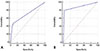
ROC curves were constructed to evaluate the diagnostic tests (sensitivity, specificity, and accuracy, based on the area under the ROC curves) for students' assessments of the PR and CBCT exams, in relation to the standard of the specialists (Fig. 3). A comparison of the values of sensitivity, specificity and accuracy through 1-way ANOVA showed that CBCT was significantly superior to PR for the diagnosis of sinus abnormalities by students (Table 4).
Discussion
The aim of the present study was to compare the efficacy of PR and CBCT for the diagnosis of maxillary sinus abnormalities by undergraduate dental students. A retrospective analysis of radiographic and tomographic images was performed, examining the maxillary sinus according to the classification proposed by Nunes et al.3 This classification was used because of its greater comprehensiveness in defining sinus abnormalities. Other classifications proposed in the literature are limited because they do not classify, for example, abnormalities such as polyps, periostitis, and antroliths,2 or only classify the origin of sinusitis without defining the type of abnormality.1
PR is a widely used diagnostic modality in routine practice at dental clinics, both by dental surgeons and undergraduate dental students,10 which justifies the importance of studies that evaluate the students' ability to analyze the accuracy of this exam in detecting abnormalities in the maxillofacial complex.
Despite its wide use, the 2-dimensional images of PR seem to play a limited role in the diagnosis of sinus abnormalities; although a broad view of the maxillary sinus is provided, the image is a flat representation of the curved surfaces of the jaws. Images of a huge number of adjacent anatomical structures are superimposed on the maxillary sinus, sometimes preventing correct interpretations of these structures.11 In addition, the radiographic signs of these abnormalities are often not specific, making it more difficult to differentiate between them in PR.12
In contrast, CBCT has emerged as the standard examination for evaluation of the maxillary sinus according to several studies in the literature.5,6,8,12,13,14 This modality provides cross-sectional images of the sinuses in different planes (sagittal, coronal, and axial), with considerable spatial resolution and high diagnostic accuracy compared to tests that provide 2-dimensional images.12 Such information explains the results found in the present study, in which PR presented reasonable agreement (kappa=0.258) in relation to the reference standard, whereas CBCT presented substantial agreement (kappa=0.692). However, according to the ROC curves, PR presented significantly lower sensitivity, specificity, and accuracy values than CBCT, meaning that it has major limitations as a diagnostic method for sinus abnormalities by students.
The present study revealed the presence of sinus abnormalities in 46.4% of the maxillary sinuses evaluated. In their sample, Tadinada et al.5 found a 72% frequency of the presence of some abnormality. However, their study was performed with a smaller sample of maxillary sinuses (n=100) and they included patients who were candidates for implant rehabilitation. Thus, such patients could have experienced the loss of dental structures due to periapical inflammatory lesions and periodontal diseases. This fact could explain the presence of a greater number of sinus abnormalities, as previous studies correlated the presence of such abnormalities with the presence of periapical and periodontal inflammatory lesions.15,16,17
Using the same classification of sinus abnormalities as in the present study, Nunes et al.3 revealed a frequency of abnormalities of 28.6%. They also showed that mucosal thickening was the most frequent abnormality, followed by sinus polyps, in agreement with the present results. However, Nunes et al.3 reported that periostitis associated with mucosal thickening was the third most frequent abnormality, differing from the findings of the present study in which periostitis was observed in only 1 maxillary sinus. That study found a correlation of sinus abnormalities with periapical abnormalities.
Maestre-Ferrín et al.2 also performed a study comparing the diagnostic accuracy of sinus abnormalities between PR and conventional CT in the digital format (Digital Imaging and Communications in Medicine image analysis in Implametric software) and images printed on film. The authors observed a frequency of normal maxillary sinuses of 96.6% on PR, 60% on conventional printed CT, and 61.6% on conventional digital CT. In the present study, there were no maxillary sinus abnormalities in 54.6% of the cases for PR, and 59.3% for CBCT. While Maestre-Ferrín et al.2 found a high number of normal sinuses on PR, the present study showed that both methods presented values close to those obtained from the reference standard for detection of normal sinuses, justifying the values (0.92 for PR and 0.97 for CBCT) and indicating that there was a low possibility of false-positive results.
When comparing the data obtained from the ROC curve with the results found by Tadinada et al.,5 it was observed that both showed higher values of accuracy for CBCT than for PR. However, Tadinada et al.5 observed a high sensitivity and low specificity for PR, unlike the present study. This divergence can be explained by the influence of the professional experience of the evaluators in the studies. In the study of Tadinada et al.,5 the evaluations were performed by dental surgeons specializing in dental radiology and periodontics.
The evaluators of the present study were undergraduate dental students and, although they were calibrated to perform the evaluations, it is important to emphasize that they were in training, with limited experience in image analysis. Gang et al.18 suggested that although observers were aware of the types of abnormalities (calibration step), a lack of experience represented a limiting factor for the accurate diagnosis of maxillary sinus abnormalities. Simuntis et al.13 reported that imaging methods for evaluation of the maxillary sinus yielded better results when the evaluators were specialists in dental radiology.
Dau et al.8 showed that professional experience in image analysis was related to the type of exam that could yield meaningful evaluation results. PR alone might not be sufficient for diagnostic evaluation by an inexperienced observer, agreeing with the results of the present study, in which PR was limited for the diagnosis of sinus abnormalities. However, when evaluated by more experienced individuals, PR was an important diagnostic tool. Different results could have been found if the evaluators in this research had more extensive clinical experience. This fact highlights the importance of teaching methodologies in the discipline of dental radiology that aim to provide more experience to students in PR interpretation, evaluating the exam in general, and diagnosing sinus abnormalities more specifically.
Although CBCT yielded significantly better results, it should be emphasized that prior to their request for CBCT to evaluate the maxillary sinuses, students should discuss the images observed in PR with more experienced professionals, who may be able to detect abnormalities not diagnosed by the students. CBCT should be requested only after a detailed investigation of the patient's clinical history and the determination that the evaluation of the PR by the student and a more experienced professional has yielded inconclusive data. This is essentially due to the need to keep the dose of radiation to the patient as low as possible for diagnosis, according to the ALADA (as low as diagnostically acceptable) principle.7
Several studies have evaluated the ability of dental students in different diagnostic tasks19,20,21 and various methodologies that promote students' cognitive development.22,23,24 Cho19 evaluated the performance of students in identifying mandibular condyle fractures, Shintaku et al.20 evaluated the ability of students to detect radiographic changes suggestive of osteoporosis, and Kratz et al.21 evaluated the interpretation of professionals in training of PR obtained from completely edentulous patients; those studies all sought to optimize the diagnosis and the training of more prepared professionals, in alignment with the objective of the present study.
In addition, Baghdady et al.22,23 and Turgeon et al.24 showed that how the issue was approached with the student also influenced the diagnosis, in addition to previous experiences and different levels of student knowledge. That is why, considering the importance of evaluating the ability of undergraduate dental students to analyze imaging exams, further studies should be performed considering different changes in the maxillofacial complex and different levels of knowledge among students. Furthermore, it is important to evaluate new teaching methodologies, seeking to improve the results obtained by learners.
In conclusion, CBCT was better than PR in detecting maxillary sinus abnormalities by dental students. However, CBCT should only be requested after a careful analysis of PR by students together with more experienced professionals.


 XML Download