

 PDF
PDF ePub
ePub Citation
 Print
Print


The jugular bulb is a confluence of the lateral dural venous sinuses that pass through the jugular foramen. The transverse and sigmoid sinuses provide the majority of the bulb's venous inflow, and the internal jugular vein provides its main outflow.12 The jugular bulb passes through the jugular foramen of the posterior cranial fossa and drains extracranially to the internal jugular vein.3 The jugular bulb is not present at birth; development of the bulb occurs in childhood, especially during the first 2 years of life. Once a child has the ability to stay upright, an erect posture causes the ascending negative pulse waves originating from the right atrium to be transmitted rostrally into the jugular sinus, leading to the dilation or formation of the jugular bulb.4 Growth of the jugular bulb continues until completion in adulthood.
The most common abnormalities of the jugular bulb are high-riding jugular bulb and jugular bulb diverticulum, the causes of which are poorly understood. Many factors are thought to impact the exact size and position of the bulb, including postnatal events, blood flow, mastoid pneumatization, and flow dynamics.2 Abnormal blood flow, whether hypertension or turbulence, is suggested as a common contributing factor to these 2 abnormalities.4 The vestibular aqueduct, facial nerve, and posterior semicircular canal may all be affected by such abnormalities.3
Jugular bulb diverticulum is an irregular extension of the jugular bulb that expands to the superior surface of the petrous bone, middle ear cavity, endolymphatic duct, or vestibular aqueduct.5 The pathophysiology of jugular bulb diverticulum formation is relatively unknown.4 Depending on its location and size, a diverticulum may be symptomatic or asymptomatic.5 In rare cases, the jugular bulb may extend into the occipital condyle; this is termed a condylar jugular diverticulum.6
This report presents a series of condylar jugular diverticula that were discovered as incidental findings on conebeam computed tomographic (CBCT) images.
Case Report
Case 1
A 60-year-old woman presented to her general dentist complaining of dental pain related to an endodontically treated right mandibular second molar. The clinician performed a CBCT scan to evaluate the right posterior mandible in an attempt to identify the source of the patient's pain. The volume of the scan extended from the level of the frontal bone to C4. The study was then referred to the Department of Oral and Maxillofacial Radiology at the University of Florida College of Dentistry (UFCOD) for radiographic interpretation of the right mandible and a general review.
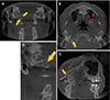
The coronal view of the CBCT scan demonstrated a well-defined corticated defect that extended medially and inferiorly from the right jugular bulb into the ipsilateral occipital condyle. No evidence of bone destruction was visible (Fig. 1A). The axial view showed that a jugular spine was maintained, and a mucus retention pseudocyst was incidentally noted in the left maxillary sinus (Fig. 1B). A sagittal CBCT image depicted an irregular expansion of the jugular bulb into the occipital condyle, as well as mucositis and a mucus retention pseudocyst in the left maxillary sinus (Fig. 1C). A sagittal image also displayed signs of a dilated jugular bulb with no mass effect on adjacent structures. The mastoid air cells appeared well-aerated (Fig. 1D). The patient had no history of jugular foramen syndrome that would suggest a space-occupying mass in the vicinity. The radiographic appearance was consistent with condylar jugular diverticulum.
Case 2
A 45-year-old woman visited her new dentist with a medical history significant for a maxillary cyst removal. The clinician performed a CBCT scan to evaluate the osseous defect and plan for maxillary and mandibular implants. The study was then referred to the Department of Oral and Maxillofacial Radiology at UFCOD for radiographic assessment of the maxilla and a general review. The volume of the maxillofacial CBCT study extended from the level of the ethmoid air cells to C4.
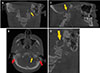
The coronal CBCT view demonstrated a well-defined corticated defect that extended medially, laterally, and inferiorly from the left jugular bulb into the ipsilateral occipital condyle. No evidence of bone destruction was visible (Fig. 2A). The axial view showed that the jugular spine was maintained, and the mastoid air cells on the left side appeared to be well-aerated compared to those on the contralateral side (Fig. 2B). A sagittal CBCT image showed irregular expansion of the jugular bulb into the occipital condyle, and reviewers also incidentally noted minimal thickening of the soft tissue of the left maxillary sinus consistent with mucositis (Fig. 2C). A sagittal image clearly showed a dilated jugular bulb extending into the occipital condyle. Adjacent structures were not impacted by any mass effect (Fig. 2D), and the patient had no history of jugular foramen syndrome. The radiographic appearance was consistent with condylar jugular diverticulum.
Case 3
A 61-year-old woman consulted a new practitioner for dental implant planning. To evaluate the maxillofacial region for implant reconstruction, the clinician performed a CBCT scan. The volume of the scan extended from the level of the frontal bone to C5. The study was then referred to the Department of Oral and Maxillofacial Radiology at College of Dentistry, University of Florida for radiographic interpretation and assistance in implant planning.
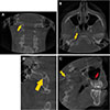
The patient presented with dental findings of periodontal bone loss, rarefying osteitis associated with multiple teeth, and radiographic results suggestive of sinusitis. The coronal CBCT view also demonstrated abnormal extension of the jugular bulb medially, laterally, and inferiorly from the right jugular bulb into the ipsilateral occipital condyle (Fig. 3A). The axial CBCT view demonstrated no evidence of bone destruction or mass effect on adjacent structures, and the mastoid air cells appeared well-aerated bilaterally. The jugular spine was maintained (Fig. 3B). A sagittal CBCT view showed an incidental finding of sinusitis in the right maxillary sinus, as well as expansion of the jugular bulb into the occipital condyle (Fig. 3C). A sagittal view also clearly showed a dilated jugular bulb extending into the occipital condyle (Fig. 3D). The radiographic appearance was consistent with condylar jugular diverticulum.
Discussion
Extension of the jugular bulb into the petrous part of the temporal bone in the superior, medial, and lateral directions is well documented. Jugular bulb extension into the temporal bone may result in pulsatile tinnitus, vertigo, or conductive hearing loss in some patients.3 However, in contrast to extension into the temporal bone, extension of the jugular bulb into the occipital condyle has rarely been reported in the literature. Condylar jugular diverticulum is a rare anatomical variant that consists of an extension of the jugular bulb into the occipital condyle.6
The occipital condyle is a distinctive bony structure, specifically a protuberance of the occipital bone, that links the skull and the vertebral column.7 The anatomical location of the jugular bulb puts it in close proximity with the occipital condyle. To the best of our knowledge, condylar jugular diverticulum has been reported only once, in the Journal of Computer Assisted Tomography in 2009, in which the author described 6 instances of a jugular bulb diverticulum extending into the occipital condyle.6 We present 3 cases of condylar jugular diverticulum extending medially and inferiorly into the occipital condyle.6 No apparent consensus exists in the literature regarding its development, and no reference describes this entity completely.
Fractures occurring near the occipital condyle can injure the area of the jugular bulb that extends into the occipital condyle, which can lead to hemorrhage in the vicinity of a diverticulum.4 Surgical skull base procedures, such as retrosigmoid craniectomy and the transcondylar-transtubular approach, are utilized to treat lesions in the middle and/or inner ear, as well as other lesions of the ventral foramen magnum and the craniovertebral junction. These surgical procedures cause extradural reduction of the occipital condyle and jugular tubercle, which can expose the hypoglossal canal and the posterior condyle and open up the posterior rim of the jugular foramen. The extension of the jugular bulb into the occipital condyle can expose or injure the jugular bulb during these surgical procedures.8
The jugular spine is a small, sharp ledge that separates the jugular foramen into the pars nervosa anteriorly and pars vascularis posteriorly. It is an important landmark, as space-occupying masses in the jugular foramen can erode the jugular spine, and asymmetry in the jugular bulb can be evaluated utilizing the jugular spine.9 The initial differential diagnosis of an asymmetrically enlarged jugular foramen may include a space-occupying mass in the jugular bulb. The presence of uniform cortication, maintenance of the jugular spine, and confluence with the jugular bulb are essential for recognition of condylar jugular diverticulum, which may be of potential significance in patients undergoing skull base surgery involving lesions in the region of the ventral medulla or foramen magnum.6
The awareness and use of CBCT in dentistry is steadily increasing.10 Given its utility in the imaging of complex head and neck anatomy, thorough interpretation of the entire CBCT scan volume is essential. Practitioners must have adequate knowledge about CBCT to ensure that they can recognize all relevant findings - including incidental and rare findings such as condylar jugular diverticulum - that could otherwise go misdiagnosed.11
In conclusion, the jugular bulb diverticulum is a wellknown anatomical variant; however, its extension into the occipital condyle has only rarely been reported in the medical literature and, to the best of our knowledge, never in the dental literature. The maintenance of the jugular spine is an important landmark to help differentiate condylar jugular diverticulum from pathosis in CBCT when soft-tissue contrast is not available. Recognition of this condylar variant may help prevent misdiagnosis when utilizing CBCT. No treatment is required for condylar jugular diverticulum.12 Exposure and injury to the jugular bulb, and possible associated complications, due to surgery in that region can be avoided through radiographic diagnosis.
 XML Download