

 PDF
PDF ePub
ePub Citation
 Print
Print


Introduction
The maxillofacial region is a common anatomical site for the development of infections, cysts, and tumors of odontogenic or non-odontogenic origin. During the evaluation of jaw swellings, in certain circumstances such as chronic inflammation, clinical examinations do not provide a complete assessment of the exact origin and nature of swellings; such cases require radiological imaging tools.12345
There are many reasons for requesting imaging information about a maxillofacial swelling, including determination of the nature of a condition, evaluating the extent of a lesion, and monitoring the progression or regression of a lesion over time. Computed tomography (CT) and magnetic resonance imaging (MRI) are recent imaging tools that are often used to clarify maxillofacial lesions' nature, extent, boundaries, and effect on the surroundings, but they remain expensive and have limitations. Ultrasonography may overcome these disadvantages of CT and MRI.67
Ultrasonography is used for the diagnosis of oral and maxillofacial swellings because it is rapid, widely available, relatively inexpensive, and painless; furthermore, it can be repeated as often as necessary without risk to the patient. In areas where a definitive diagnosis cannot be established, ultrasonographic features can at least be used to categorize the swelling type. This directive analysis can justify the further investigations required and help initiate the appropriate treatment plan. Although many studies have investigated correlations between ultrasonographic features and maxillofacial lesions, little research has sought to classify maxillofacial lesions according to ultrasonographic features. Therefore, this study aimed to evaluate various features appearing on ultrasonographic examinations of maxillofacial lesions.89101112
Materials and Methods
Fifty patients with swellings in the oral and/or maxillofacial region were randomly selected from the outpatient clinics of Minia University Hospital, Minia University Dental Hospital, and Minia General Hospital. Their minimum age and maximum age were recorded, with standard deviations. Swellings caused by trauma and/or fracture and those that extended below the neck were excluded from this study. This study was approved by the Research Ethics Committee (REC), Faculty of Dentistry, Minia University before starting the research, and all patients signed a standardized informed consent form established by the REC.
A comprehensive questionnaire was used to assess case history, and thorough extra-oral and intra-oral examinations were then carried out. The ultrasonographic investigations were carried out at the Department of Radiology of Minia University Hospital using an ultrasound diagnostic modality (LOGIQ-P5) (GE Medical System, Seongnam, Korea) with color Doppler function with a linear array transducer, operating at a frequency of 7.5–12 MHz. All examinations were performed over the swellings and compared to the contralateral/normal side when appropriate. All ultrasonographic images were interpreted by an expert ultrasonologist (15 years' experience). Ultrasonographic features were then recorded according to the characteristics reported by Shimizu et al.,13 including shape, boundary, echo intensity, ultrasound architecture of the lesion, posterior echoes, ultrasound architecture of the tissues, vascularity, presence of necrosis, and presence of calcification. The ultrasonography-guided diagnosis was obtained according to these features, and the lesions were initially categorized into 5 groups for histopathology. Finally, patients underwent either fine needle aspiration or biopsy for a histopathological examination to obtain the final diagnosis. However, in inflammatory swellings, the final diagnosis was established by a blood panel and the response of the swelling to either surgical intervention (i.e., incision and drainage) or successful medical/non-surgical treatment. According to the final diagnosis, the patients' swellings were categorized into 5 groups; group I: inflammatory/infection/abscess swellings, group II: cystic swellings, group III: lymph node swellings, group IV: benign swellings, and group V: malignant neoplastic swellings.
The associations between the ultrasonography-guided diagnosis and histopathological findings and the contingency coefficient were analyzed using SPSS version 20 (IBM Corp., Armonk, NY, USA). The same software was used to calculate the sensitivity, specificity, positive predictive value (PPV), negative predictive value (NPV) and accuracy of the ultrasonography-guided diagnoses, using the histopathological findings as a reference. P values were calculated using the chi-square test in all study groups.
Results
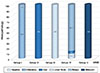
Among the 50 studied individuals, there were 17 males (34%) and 33 females (66%) who had oral and/or maxillofacial swellings. The minimum age was 8 years and the maximum was 71 years, with a mean age±standard deviation of 35.3±17.1 years. A highly significant association was observed between the ultrasonographic and histopathologic diagnoses with a contingency coefficient of 0.88 and a P value<0.05. This association reached 100% in groups III and V, followed by 94.7% in group IV and 91% in groups I and II (Fig. 1).
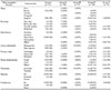
According to Table 1, most inflammatory swellings were finally diagnosed as cellulitis, dento-alveolar abscesses, space infections, osteomyelitis, or Ludwig angina. In more detail, 82% had irregular shapes, 91% showed hypoechoic echogenicity, 82% displayed heterogeneous ultrasonographic architecture of the lesion, and 63% exhibited posterior enhancement. The cystic swellings (group II) were finally diagnosed as 4 radicular cysts, 2 dentigerous cysts, 2 nasolabial cysts, 1 epidermoid cyst, and 1 calcifying epithelial odontogenic cyst. Of these cysts, 70% had very clear boundaries, 80% showed homogenous lesion architecture, 90% displayed enhanced posterior echoes and 100% had neither vascularity nor necrosis. Group III was finally diagnosed as 3 lymph node abscesses, 1 benign lymphadenitis, and 1 metastatic lymph node. Of the lymph node abscesses, 60% had ill-defined boundaries, 100% showed homogenous lesion architecture, 80% had cystic tissue characteristics, 60% displayed posterior enhancement, and 80% had central necrosis and calcifications; moreover, vascularity was detected in 0% of these lesions. On ultrasonography, abnormal benign lymph nodes appeared as hypoechoic or mixed echogenicity with the reversible loss of the central hilus and an increase in the short axis measurement. In contrast, in malignant nodes (20%), destruction of the central fatty hilum was evident and the lymph nodes were more oval with anechoic echogenicity.
The benign swellings (group IV) were histopathologically diagnosed as 1 ossifying fibroma, 3 cases of sialolithiasis, 3 pleomorphic adenomas, 2 brown tumors of hyperparathyroidism, 2 ameloblastomas, 1 central giant cell granuloma, 1 oral ranula, 1 case of Sjögren syndrome, 1 arteriovenous malformation, 1 adenomatoid odontogenic tumor, 1 fibroma, 1 neurofibromatosis, 1 bony exostosis (torus mandibularis), 1 fibrous dysplasia, and 1 cavernous hemangioma. Of these lesions, 67% showed heterogenous ultrasound architecture of the lesion, 57% had mixed tissue characteristics, 67% were without vascularity, 90% were without necrosis, and 81% were without calcifications. The malignant neoplastic swellings (group V) included 2 squamous cell carcinomas and 1 rhabdomyosarcoma, as confirmed by histopathology. Of these lesions, 100% had irregular shapes, 100% showed heterogenous ultrasound architecture of the lesion, 100% were without vascularity, and 100% were without calcifications (Table 1).
Table 2 shows that the ultrasonographic diagnostic accuracy was 100% in lymph node and malignant swellings, followed by 98% in inflammatory and cystic swellings and 92% in benign swellings, with a total diagnostic accuracy of 89%. The sensitivity of the ultrasonography-guided diagnoses was 100% in cystic, lymph node, and malignant swellings, followed by 91% in inflammatory swellings and 86% in benign swellings.
Discussion
Ultrasonography was used in this study for the diagnosis of oral and maxillofacial swellings because it is a valuable aid to oral and maxillofacial surgeons, as it is rapid, widely available, relatively inexpensive, and painless; furthermore, it can be repeated as often as necessary without risk to the patient. The ultrasonographic features could be used to categorize the swelling type, which can help to initiate the appropriate treatment plan. Furthermore, ultrasonography could provide an alternative to radiography, especially in unilocular jaw bone lesions that are difficult to diagnose because of their similar radiographic appearance.89101112
In the present study, swellings owing to trauma or fracture were not included because the provisional diagnosis of hematoma can be straightforwardly made on the basis of a history of trauma and changes in skin color and the mucous membrane.
In this study, the minimum age of the selected 50 individuals was 8 years and the maximum was 71 years, with a mean age±standard deviation of 35.3±17.1 years. This corresponds well with another study, in which there were 30 subjects with minimum and maximum ages were of 8 years and 65 years, respectively, and a mean age±standard deviation of 33.13±5.36.9
In the present study, the diagnostic accuracy of ultrasonography in diagnosing maxillofacial swellings was 89% when compared to histopathology. This agrees with the results of other researchers, who reported that the diagnostic accuracy of ultrasonography in maxillofacial swellings was 88.9% in comparison to the histopathological diagnosis.
In addition, the accuracy of ultrasonography in detecting inflammatory, cystic, lymph node, benign, and malignant swellings was 98%, 98%, 100%, 92%, and 100%, respectively. For comparison, another study reported that the congruence of ultrasonography with histopathology was 78% in abscesses and infections, 100% in lymphadenitis, 75% in malignancies, 100% in cystic tumors, and 88% in benign tumors.2
In this study, the diagnostic accuracy of ultrasonography was 98% for inflammatory swellings. This agrees with a previous study reporting that out of 10 inflammatory swellings, which included 3 cases of osteomyelitis and 7 cases of space infections and abscesses, 9 cases were correctly identified by ultrasound, giving a diagnostic accuracy of 90%.2314
In this study, 1 swelling was misdiagnosed by ultrasound as fibrous dysplasia, but was confirmed by histopathology to be chronic suppurative osteomyelitis. This might have been due to inaccurate ultrasonographic features that appeared from the bone infection, as it had a relatively clear boundary with mixed echogenicity and mixed tissue character, in addition to the similar features of both lesions.
This study found that the characteristic ultrasonographic features of most inflammatory swellings involved irregular shapes, with ill-defined boundaries, hypoechoic echogenicity, and heterogenous ultrasound architecture of the lesion. This is in agreement with other studies reporting that inflammatory swellings were characterized by irregular shapes, hypoechoic echogenicity, and enhanced posterior enhancement. However, some researchers have stated that the boundaries of inflammatory swellings were relatively clear, with homogenous lesion architecture.10
Moreover, 82% of inflammatory lesions in this study did not appear on Doppler examinations, in contrast with other studies that reported that abscesses appeared to have a vascular supply on color Doppler examinations.1516
In this study, the accuracy of ultrasonography for detecting cystic lesions was 98%, with 91% sensitivity. These findings agreed with other studies reporting that the sensitivity and accuracy of ultrasonography for detecting cystic lesions were 92%.21718
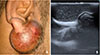
In group II, the ultrasonographic features of most cystic swellings included very clear boundaries, oval or round shapes, and mixed echogenicity (Fig. 2). This agrees with previous research findings that most cystic swellings had very clear boundaries, round shapes, and anechoic echogenicity. Other studies stated that odontogenic keratocytes were hypoechoic because of their dense content, while others reported that radicular cysts appeared as anechoic to hypoechoic, well-contoured cavities surrounded by bony walls, filled with fluid and with no evidence of internal vascularization on color Doppler examinations.291718
In this study, 1 case was diagnosed by histopathology as plexiform ameloblastoma, but it was misdiagnosed by ultrasonography as a dentigerous cyst; this could have occurred because ameloblastoma might develop from a long-standing dentigerous cyst associated with an impacted tooth. In addition to their potential to attain a large size, follicular cysts are noteworthy for their tendency to develop neoplastic changes, such as plexiform ameloblastoma and carcinoma, within an isolated segment of the cyst wall.6
In a study conducted by Pallagatti et al. in 2012, of 13 cases that were diagnosed by histopathology as 3 odontogenic keratocytes, 1 dentigerous cyst, and 9 radicular cysts, ultrasonography identified 12 cysts correctly. That finding is compatible with those of the present study, and could be explained by the specific features of the cysts on ultrasonographic examinations.2
In this study, ultrasonography detected all lymph node swellings accurately with 100% diagnostic accuracy, harmonizing with other finding that ultrasonography showed anaccuracy of 100% in detecting lymph node swellings.19
Another study concluded that ultrasonography could identify 1 case of benign lymphadenitis and 2 metastatic cervical lymph nodes confirmed by histopathology. It reported 100% diagnostic accuracy of ultrasonographic examinations in detecting benign lymphadenitis and metastatic lymph nodes.2
In group III, the characteristic ultrasonographic features of abnormal benign lymph nodes were hypoechoicity or mixed echogenicity, ill-defined boundaries, and reversible loss of the central hilum in addition to central necrosis. Reversible loss of the lymph node hilum (appearing as mixed echogenicity on ultrasonography) might be due to pus spread in a node that returned to normal echogenicity when drained. In malignant nodes, the destroyed central hilum (appearing anechoic on ultrasound) did not return to normal echogenicity and had no evidence of necrosis. This agrees with another study that concluded that abnormal nodes were hypoechoic on ultrasonography with loss of the central hilum. With ultrasonography, physicians can evaluate important parameters such as lymph node shape, margins, internal structure, and abnormal vascularization.2
The diagnostic accuracy of ultrasonography in diagnosing different benign swellings was found to be 92%, with 86% sensitivity and 96.6% specificity. Other authors reported a 100% sensitivity of ultrasonography for diagnosing solid tumors, while others reported that ultrasonography could characterize the flow of blood and differentiate hemangiomas from other lesions.217
The differences in the accuracy of detecting benign tumors in this study from other studies could be attributed to multiple factors. Two cases could not be identified. The first was a submental salivary gland stone (sialolithiasis) and the second was a benign bony exostosis in the mandibular premolar-molar area (torus mandibularis). The presence of a cortical bony plate overlying the swelling did not allow the penetration of sound waves and made it difficult to visualize the internal structure of the swelling. Hence, in some cases, it was observed that the efficacy of imaging could be limited due to anatomical considerations, and/or overlapping features of benign pathologies.2021
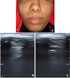
In group IV of this study, most studied benign neoplasms had very clear boundaries, irregular shapes, and hypoechoic echogenicity. The ultrasound architecture of benign neoplasms was homogeneous, with enhanced posterior echoes and mixed ultrasound characteristics of tissues (Fig. 3). These findings are similar to those of another study that reported the same ultrasonographic features of benign swellings.11
The study of Bhardwaj et al.11 found a benign mass lesion in the left maxilla, which demonstrated a hypoechoic internal echo pattern with areas of calcifications and was histopathologically diagnosed as desmoplastic ameloblastoma. Two other cases were diagnosed histopathologically as lipomas, and showed a hypoechoic internal echo pattern with homogenous echoes on ultrasonographic examinations.
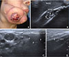
Group V constituted 3 cases, which were diagnosed by histopathology as 2 squamous cell carcinomas and 1 rhabdomyosarcoma (Fig. 4). Ultrasonography identified all of them correctly with 100% diagnostic accuracy, while the diagnostic accuracy of ultrasonography in detecting malignancies in other studies was 82%. Another study stated that the diagnostic accuracy of ultrasonography in differentiating benign and malignant lesions was 67%, in contrast to this study and other studies that reported accuracy to be as high as 80–88%.2223
In this study, the most studied malignant neoplasms had mixed echogenicity, while other authors concluded that malignant tumors showed a complex echotexture with a heterogeneous internal echo pattern and irregular boundaries.10222425
In conclusion, ultrasonography is a recommended imaging tool for diagnosing maxillofacial swellings. Ultrasonographic features together with Doppler function greatly aid in making an accurate diagnosis of an oral and/or maxillofacial swelling. If the ultrasonographic features of an oral and/or maxillofacial lesion include an irregular shape, hypoechoicity, and heterogenous lesion architecture, it is likely to be an inflammatory or infected lesion. The features of very clear boundaries, homogenous lesion architecture, and enhanced posterior enhancement are suggestive of cystic lesions. Lymph node features include ill-defined boundaries, homogenous lesion architecture, and central necrosis. Ultrasonographic features of heterogenous lesion architecture, with mixed tissue characteristics and enhanced posterior echoes, are suggestive of benign lesions. However, irregular lesion shapes, with heterogenous lesion architecture and eccentric necrosis, suggest a malignant neoplasm.

 XML Download