

 PDF
PDF ePub
ePub Citation
 Print
Print


Dear Editor:
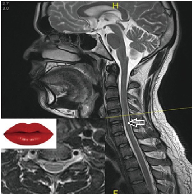
We would like to highlight an important sign that is observed during magnetic resonance imaging (MRI) of the spinal cord, and is designated by us as the “lip sign” owing to its typical appearance. This sign, observed in the axial T2-weighted (T2W) MRI sequence of the spinal cord, is secondary to truncation artifacts (Gibbs phenomenon), and not due to any underlying pathology. It maybe an important sign to observe, as often times these artifacts can be mistaken for other pathologies such as syrinx, demyelination, or persistent central canal (1).
Artifacts in MRI are generally caused by either the scanner hardware itself or the interaction between the patient and MRI scanner (1). Truncation signal artifacts appear as ripple features in areas where there are sudden transitions between low and high signal intensity (2). This particular artifact is generally found at the interface between the spinal cord, which is of low signal intensity, and the cerebrospinal fluid, which is of high signal intensity, on the sagittal and axial T2W MRI sequences of the spine (34). It may be observed as a hyperintense line that runs parallel along the dorsal and ventral regions of the spinal cord on the T2W sequence (5). The main physics behind such artifacts is that, when the magnetic resonance signal is sampled over a limited period, some of the data is usually omitted or truncated in the k-space, causing the signal intensity of a given pixel on the final image to differ from the true signal intensity (4).
This truncation artifact has a typical appearance of ‘lips’ in the axial T2W MRI sequence of the spine, and is also typically observed in the cervical and thoracic spinal cord of patients in any age group, with both 1.5T and 3T MRI scanners. Thus, if this abnormality is observed in a sagittal T2W scan of the spine, it becomes pertinent to check the axial section at the same level; the presence of the ‘lip sign’ in an axial scan denotes it to be an artifact (Fig. 1). The presence of this artifacthere was confirmed by normal post-contrast scan, while diffusion tensor imaging of the spine was also performed, which showed no significant difference in fractional anisotropy values in the region of the spine with abnormal T2W signal changes, relative to the region where no abnormal signal change was seen. Furthermore, the diffusion-weighted imaging sequence also showed no abnormal diffusion restriction in the area of the abnormal signal.
The sole purpose of this article was not to provide a detailed description of the “truncation artifact,” which is a concept already discussed in many articles, but to describe a sign that has not been mentioned previously in any related studies, that may lead to a confident diagnosis of this artifact. This, in turn, will avoid an increase in scan time by excluding additional sequences and unnecessary follow-up scans in such cases, thus preventing an additional cost of procedure and unnecessary mental stress to patients and their relatives, concerning a pathology that is actually not present.
 XML Download