

 PDF
PDF ePub
ePub Citation
 Print
Print


INTRODUCTION
Thyroid core needle biopsy (CNB) has been proposed as an additional diagnostic method to ultrasound (US)-guided fine needle aspiration (FNA), mainly to overcome the limitations of nondiagnostic or inconclusive cytologic diagnosis, which can lead to repeat FNAs or unnecessary surgery (12). In addition, thyroid CNB has been recently applied to various indications, including the differentiation of rapidly growing thyroid tumors, differentiation of follicular lesions, medullary thyroid carcinoma, calcified thyroid nodules, and degenerating thyroid nodules (345). Furthermore, several studies have even suggested the value of CNB as a first-line diagnostic tool for the thyroid (6789).
In 2016, the Korean Society of Thyroid Radiology revised the consensus statement and recommendations for CNB of thyroid nodules (5). Although these guidelines include 11 recommendations regarding the indications, device, procedure, clinical outcomes, and complications, an obvious CNB protocol has not been clearly established yet. Previously, we suggested the modified CNB technique which targets nodular tissue, the nodular margin, and the surrounding parenchyma simultaneously (i.e., marginal target) (10). In our later study that used this modified technique, the CNB protocol for cytologically inconclusive thyroid nodules required at least two specimens with both intranodular and marginal targets (11). Although this protocol was not based on the US findings, recent studies by Ahn et al. (12) and Kim et al. (13) suggest that a modified CNB technique may be useful in the diagnosis of thyroid nodules with specific US features.
In the present study, we attempted to modify our previously proposed CNB protocol to improve its value in clinical application. Therefore, the purpose of this study was to retrospectively compare the diagnostic performances of two different biopsy techniques for obtaining two core specimens, either by marginal only or marginal with intranodular targets, for thyroid nodules with low or intermediate suspicion US patterns.
Go to : 
MATERIALS AND METHODS
The Institutional Review Board of Samsung Medical Center, Sungkyunkwan University School of Medicine, approved this retrospective study and waived the informed consent requirement. However, all US-guided biopsies were conducted after obtaining informed consent from the patients.
Patients
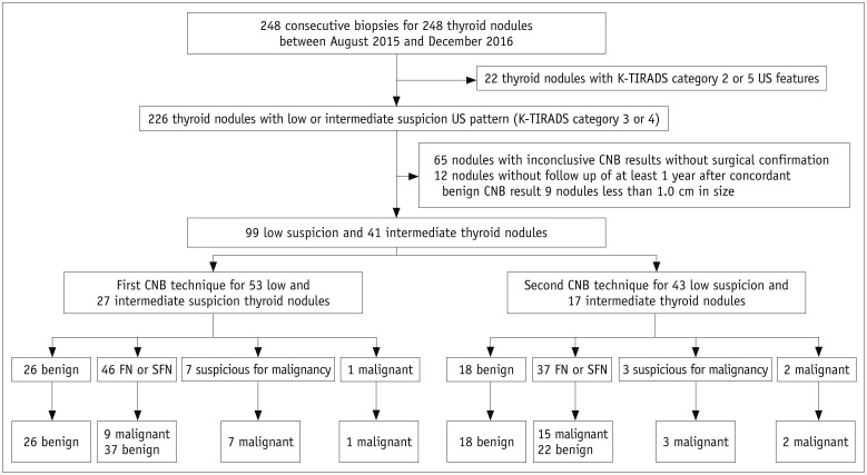
From August 2015 to December 2016, a total of 248 consecutive CNBs for 248 thyroid nodules were performed at our institution. During the period, CNB was used as a first-line biopsy or as a repeat biopsy for nodules with previous inconclusive FNA results. We retrospectively included Korean Thyroid Imaging Reporting and Data System (K-TIRADS) category 4 or 3 nodules with sizes of 1 cm or larger and final diagnoses in this analysis. A final diagnosis of malignancy was made based on surgical pathology. A final diagnosis of benign nodule was made when one of the following conditions was met: surgical diagnosis, concordant benign results after biopsy on at least two occasions, or an initial result of benign biopsy and decreased or stable size at follow-up US more than 1 year later. Then, we excluded 77 nodules without final diagnoses (inconclusive CNB results without surgical confirmation [n = 65] or lack of follow-up of at least 1 year after a concordant benign CNB result [n = 12]); 22 nodules of K-TIRADS categories 2 or 5; and 9 nodules smaller than 1.0 cm in size. Finally, the study included 135 patients (98 women and 37 men; mean age, 48.0 ± 12.4 years) with 41 intermediate and 99 low suspicion thyroid nodules (i.e., K-TIRADS category 4 or 3) (Fig. 1).
Ultrasonography and Ultrasonography-Guided Core Needle Biopsy Procedures
All thyroid nodules were examined using a 7–12 MHz linear transducer (iU22, Philips Medical Systems, Bothell, WA, USA). The US images were retrospectively reviewed in consensus according to the K-TIRADS by two board-certified radiologists who specialize in thyroid imaging with more than 8 years of experience. The K-TIRADS category was decided as high suspicion (5), intermediate suspicion (4), low suspicion (3), or benign (2) nodule based on the malignancy risk stratified by US patterns composed of integrated solidity, echogenicity, presence of microcalcification, orientation, and margin (14). According to the K-TIRADS, category 3 represents a low suspicion nodule (partially cystic or isohyperechoic nodule without any of the three suspicious US features, with a 3–15% malignancy risk) and category 4, an intermediate suspicion nodule (solid hypoechoic nodule without any of the three suspicious US features or partially cystic or isohyperechoic nodule with any of the three suspicious US features, with a 15–50% malignancy risk).
US-guided CNB was performed by one of the two board-certified radiologists who specialize in thyroid imaging with more than 8 years of experience, using a disposable 18-gauge, double-action, spring-activated needle (11 mm penetration with a 7 mm sample notch) (TSK Ace-cut, Create Medic, Yokohama, Japan). With reference to our previous study (11), we routinely obtained two specimens for thyroid core biopsies since August 2015. In particular, throughout this study period, two different biopsy techniques were alternatively applied to the consecutive thyroid nodules. During the study period, the first and second biopsy techniques were initially performed for each of the 124 cases. Based on our exclusion criteria, 44 cases with the first technique and 64 cases with the second technique were excluded from the final population (Fig. 2). In the first technique, the two specimens were targeted to include the nodular tissue, nodular margin, and the surrounding normal parenchyma simultaneously (i.e., marginal target) (10). In the second technique, the two specimens were obtained from two different target areas, one from the marginal target and another from the intranodular target. With the second technique, each biopsy specimen was placed in a separate container which contained formaldehyde solution and was labeled for identification of the different biopsy sites. All specimens were submitted for routine pathologic examination.
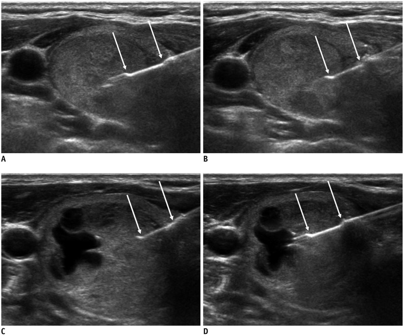 | Fig. 2Two CNB techniques.In first biopsy technique (A, B), two specimens included nodular tissue, nodular margin, and surrounding normal parenchyma (i.e., margin target). In second technique (C, D), two specimens were obtained from two different target areas, one from marginal target (C) and another from intranodular target (D). Arrows indicate specimen notch of biopsy needle.
|
Histologic Analysis
All biopsy specimens were mounted on separate slides and retrospectively reviewed by one experienced pathologist who was blinded to the original pathology report.
The CNB results were assigned to 1 of 6 categories, according to the research of Jung et al. (15). According to this reporting system, category I corresponds to nondiagnostic or unsatisfactory; category II, benign lesion; category III, indeterminate lesion (indeterminate follicular lesion with nuclear atypia/architectural atypia); category IV, follicular neoplasm or suspicious for follicular neoplasm; category V, suspicious for malignancy; and category VI, malignancy. Then, all thyroid nodules were divided into two subgroups: non-neoplasm (CNB categories I, II, and III) versus neoplasm (CNB categories IV, V, and VI), and malignancy (CNB categories V and VI) versus benign nodules (CNB categories I, II, III, and IV).
Statistical Analysis
Statistical analysis was performed using SPSS, version 23 (IBM Corp., Armonk, NY, USA). For diagnosing thyroid neoplasm and malignancy, we calculated the sensitivity, specificity, positive predictive value (PPV), negative predictive value (NPV), and the accuracy of CNB. Chi-square and Fisher's exact tests were used to compare the CNB and final diagnoses between the two different biopsy technique groups and to compare the diagnostic performances of the two different biopsy techniques to predict thyroid neoplasm and malignancy. A statistically significant difference was defined as p < 0.05.
Go to :
RESULTS
During the study period, CNB was performed for 80 intermediate or low suspicion thyroid nodules (57.1%) using the first technique (marginal targets only), and 60 nodules (42.9%) using the second technique (marginal and intranodular targets) (Table 1). Between the two groups with different biopsy techniques, there were no significant differences in the age and sex of patients, as well as the size and K-TIRADS category of the thyroid nodules.
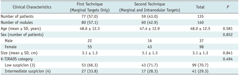
Table 1
Demographic Data of Patients with 140 Thyroid Nodules
![]()
Table 2 presents the CNB data and final diagnoses according to the biopsy technique. Between the two biopsy techniques, there were no significant differences in the CNB diagnoses (p = 0.686). According to the final diagnosis, the malignancy rate was 21.2% (17/80) in the first technique group and 33.3% (20/60) in the second technique group (p = 0.177). There were no CNB category I or III diagnoses in this population. For predicting thyroid malignancy, neither technique demonstrated false-positive results in the CNB category V and VI nodules. In addition, neither technique produced a false-negative result in the CNB category II nodules. Among the CNB category IV nodules, false-negative diagnosis was found in nine nodules (five follicular variant of papillary thyroid carcinoma [FVPTC], three follicular thyroid carcinoma [FTC], and one classic PTC) using the first technique and 15 nodules (nine FTC, five FVPTC, and one classic PTC) using the second technique. For predicting thyroid neoplasm, the first biopsy technique produced neither false-positive nor false-negative diagnoses, while there were three false-negatives (all follicular adenomas) among the CNB category II nodules and one false-positive (benign nodule) CNB category IV nodule.
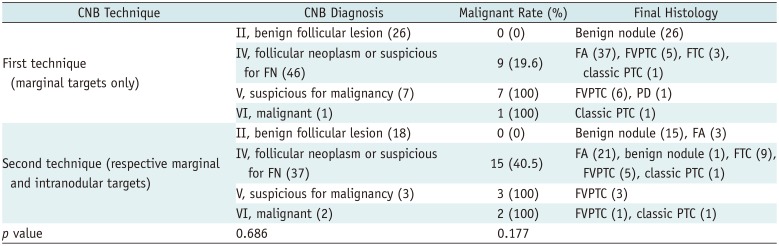
Table 2
Data of CNB and Final Diagnoses according to Biopsy Technique
Numbers in parentheses indicate number of lesions. CNB = core needle biopsy, FA = follicular adenoma, FN = follicular neoplasm, FTC = follicular thyroid carcinoma, FVPTC = follicular variant of papillary thyroid carcinoma, PD = poorly differentiated thyroid carcinoma, PTC = papillary thyroid carcinoma
![]()
Table 3 summarizes the diagnostic performance of the two different CNB techniques for predicting thyroid neoplasm and malignancy. The accuracy of the first biopsy technique was significantly higher than that of the second biopsy technique (100% vs. 93.3%, p = 0.032 for predicting thyroid neoplasm; 88.8% vs. 75.0%, p = 0.033 for predicting thyroid malignancy). The NPV of the first biopsy technique for predicting thyroid malignancy was significantly higher than that of the second biopsy technique (87.5% vs. 72.7%, p = 0.035). The sensitivity, specificity, and PPV were comparable between the two different biopsy techniques.
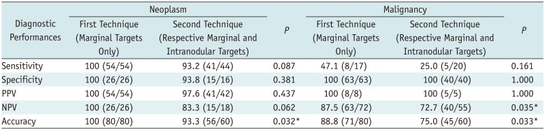
Table 3
Diagnostic Performances of Two CNB Techniques for Thyroid Neoplasm and Malignancy
![]()
In our study, one patient had subsequent perinodular hemorrhage after the first technique and another had minimal intra-parenchymal hemorrhage after the second technique, both of which were resolved by manual compression (1.4%).
Go to :
DISCUSSION
We previously published results describing the ideal core number for US-guided thyroid biopsy of cytologically inconclusive nodules (11). Our results indicated that US-guided CNB for cytologically inconclusive nodules should obtain at least two cores with intranodular and additional marginal targets. Since completing that study, we routinely obtained two core specimens during thyroid biopsy at our institution. During the study period, the first and second biopsy techniques were alternatively performed for each of the 124 cases by one of the two experienced radiologists. As described above, two core specimens were obtained targeting the margin only (i.e., the first technique) or targeting the intranodular and marginal tissue (i.e., the second technique, the same protocol proposed in the previous study) (11). The current results indicate that the first technique is better than the second technique for predicting thyroid neoplasm and malignancy. Therefore, thyroid core biopsy should obtain at least two cores with marginal targets, particularly for intermediate or low suspicion thyroid nodules.
K-TIRADS category 5, high suspicion nodules, are usually considered classic PTC (1416). Because classic PTC is diagnosed based on their nuclear features (17), most of them can be easily diagnosed by FNA. Therefore, even in the case of performing CNB, classic PTC is usually easily diagnosed from a single biopsy specimen if it is well targeted (intranodular target) (11). However, intranodular tissue retrieved by CNB is sometimes insufficient for differential diagnosis of follicular proliferative lesions because the presence of the capsule around the nodule is a major difference between nodular hyperplasia and follicular neoplasm (171819). Therefore, tissue retrieval with a marginal target that includes the nodular tissue, nodular margins, and surrounding normal parenchyma is important for differentiating a follicular neoplasm from a non-neoplastic lesion (10). We introduced this theory-based technique as a “modified CNB technique” in our previous study (10). In the present study, we demonstrated that CNB using marginal targets only (i.e., modified CNB technique only) was more effective for diagnosing cytologically inconclusive thyroid nodules with low or intermediate suspicion US patterns, compared to CNB with intranodular and additional marginal targets. On US, a large proportion of the follicular proliferative lesions, even follicular carcinomas, show low or intermediate suspicion US patterns and are finally categorized as K-TIRADS category 3 or 4 (2021222324).
Low or intermediate suspicion US patterns corresponding to K-TIRADS category 3 or 4 are predominant imaging features of FVPTCs and follicular proliferative lesions, such as nodular hyperplasia, follicular adenoma, and follicular carcinoma (2021222324). In this study, we found 61 follicular adenomas (43.6%), 42 benign nodules (30.0%), 20 FVPTCs (14.3%), 12 follicular carcinomas (8.6%), four PTCs (2.9%), and one poorly differentiated thyroid carcinoma (0.7%). For the four PTCs and one poorly differentiated thyroid carcinoma, the K-TIRADS category was “intermediate suspicion, 4.”
Although CNB performed by experienced radiologists is safe and well-tolerated, there are still safety concerns (252627). The complication rate in our study was 1.4%, similar to that in other studies, ranging from 0% to 1.5% (2829). To minimize the potential for complications, we routinely used color Doppler US when we decided the appropriate biopsy route and immediately applied manual compression on the biopsy site for at least 20 minutes after each biopsy procedure.
Our study has several limitations. First, this study was not completely randomized, although we tried to apply the two different biopsy techniques alternatively to the consecutive thyroid nodules during the study period. However, in our study, no statistical differences were found in the age and sex of patients, the size and K-TIRADS category of thyroid nodules, and the CNB diagnosis between the two different biopsy techniques. Second, the accuracy of CNB depends on several factors, such as the operators' skills and experience. However, inherent nodule characteristics can be more important factors in inducing unsatisfactory sampling under US guidance, compared to operator factors (3031). In this study, we included 140 thyroid nodules with relatively similar characteristics of K-TIRADS category 3 or 4. We believe that this inclusion criterion made it possible to include patients with relatively uniform inherent nodule characteristics. Third, in this study, we used an 11-mm excursion core device which retrieves a core specimen of approximately 0.7 cm in length. If we used a 16- or 22-mm excursion core device, one core specimen including nodular tissue, nodular margin, and surrounding normal parenchyma, could be enough for diagnosis.
In conclusion, for intermediate or low suspicion thyroid nodules, US-guided core biopsy to obtain two specimens with marginal targets only is more effective for diagnosing thyroid neoplasm or malignancy than CNB with respective marginal and intranodular targets.
Go to :
 XML Download