

 PDF
PDF ePub
ePub Citation
 Print
Print


Abstract
Metastasis to the pituitary gland is unusual, and metastasis to pituitary adenoma is extremely rare. We report here on a case of hepatocellular carcinoma metastasizing to a pituitary adenoma with MRI findings.
References
1. He W, Chen F, Dalm B, Kirby PA, Greenlee JD. Metastatic involvement of the pituitary gland: a systematic review with pooled individual patient data analysis.Pitu/iitary. 2015; 18:159–168.
2. Wilson TC, Kirby PA. A 50-year-old man with back pain and a sellar mass. Metastatic hepatocellular carcinoma. Brain Pathol. 2013; 23:365–366.
3. Bret P, Jouvet A, Madarassy G, Guyotat J, Trouillas J. Visceral cancer metastasis to pituitary adenoma: report of two cases. Surg Neurol. 2001; 55:284–290.

4. Hanna FW, Williams OM, Davies JS, Dawson T, Neal J, Scanlon MF. Pituitary apoplexy following metastasis of bronchogenic adenocarcinoma to a prolactinoma.Clin Endocrinol (Oxf). 1999; 51:377–381.
5. Noga C, Prayson RA, Kowalski R, Sweeney PJ, Mayberg M. Metastatic adenocarcinoma to a pituitary adenoma. Ann Diagn Pathol. 2001; 5:354–360.
6. Nassiri F, Cusimano M, Rotondo F, Horvath E, Kovacs K. Neuroendocrine tumor of unknown origin metastasizing to a growth hormone-secreting pituitary adenoma.World Neurosurg. 2012; 77:201.e9–201. .e12.
7. Yang C, Liu L, Lan X, Zhang S, Li X, Zhang B. Progressive visual disturbance and enlarging prolactinoma caused by melanoma metastasis: a case report and literature review. Medicine (Baltimore). 2017; 96:e6483.
8. Habu M, Tokimura H, Hirano H, Yasuda S, Nagatomo Y, Iwai Y, et al. Pituitary metastases: current practice in Japan. J Neurosurg. 2015; 123:998–1007.
9. Tanaka T, Hiramatsu K, Nosaka T, Saito Y, Naito T, Takahashi K, et al. Pituitary metastasis of hepatocellular carcinoma presenting with panhypopituitarism: a case report. BMC Cancer. 2015; 15:863.
10. Gopan T, Toms SA, Prayson RA, Suh JH, Hamrahian AH, Weil RJ. Symptomatic pituitary metastases from renal cell carcinoma.Pituitary. 2007; 10:251–259.
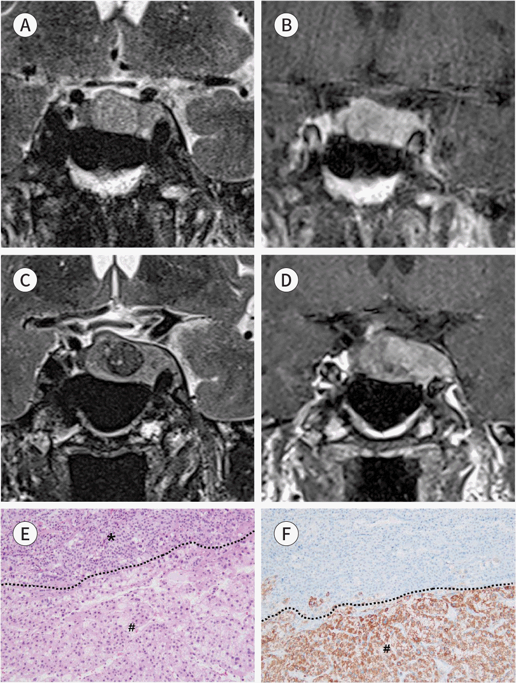
Fig. 1.
A 72-year-old man with HCC metastasis to pituitary adenoma presenting with oculomotor nerve palsy. A, B. Initial sella MRI (A, coronal T2-weighted; B, coronal contrast-enhanced T1-weighted image 30 second after administration of gadolinium) demonstrate a tumor (2.6 × 1.1 cm) located in pituitary fossa and left cavernous sinus. The tumor is revealed as slight hyper-intensity compared with gray matter on T2-weighted image and relatively homogeneous enhancement after administration of gadolinium. C, D. Two years later, sella MRI (C, coronal T2-weighted; D, coronal contrast-enhanced T1-weighted image 30 second after administration of gadolinium) show increase in size of pituitary tumor (2.8 × 1.3 cm). On T2-weighted image (C), distinct hypointense nodular area is seen within pre-existing sellar tumor showing strong enhancement after admis-tration of gadolinium on T1-weighted image (D). E, F. Histological tumor specimens (× 200). (E) Hematoxylin and eosin staining of the pituitary tumor shows metastatic HCC (#) to pituitary adenoma (∗). (F) Hepatocyte specific antigen immunohistochemical result shows reactive in metastatic HCC (#). HCC = hepatocellular carcinoma
 XML Download