Abstract
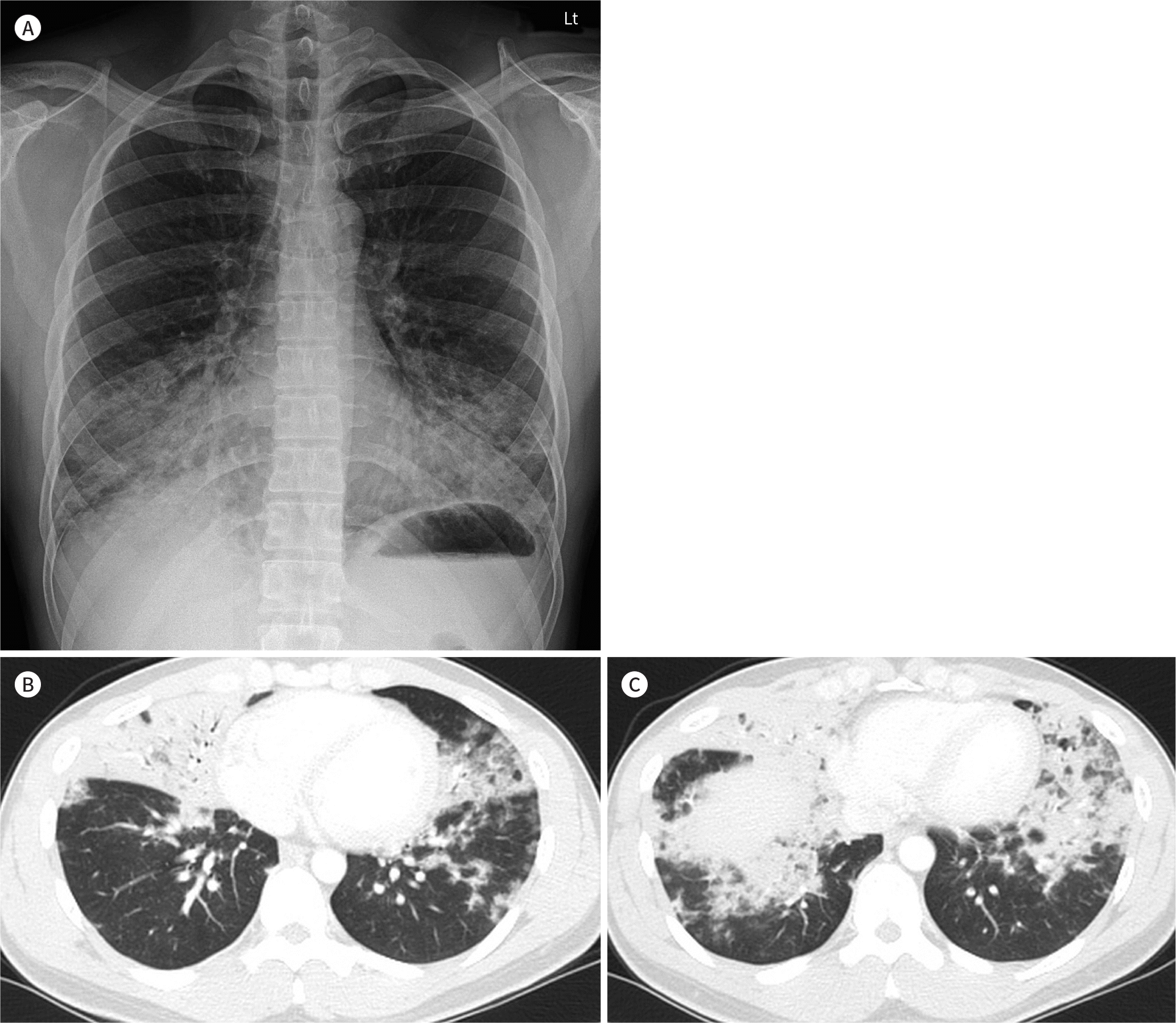
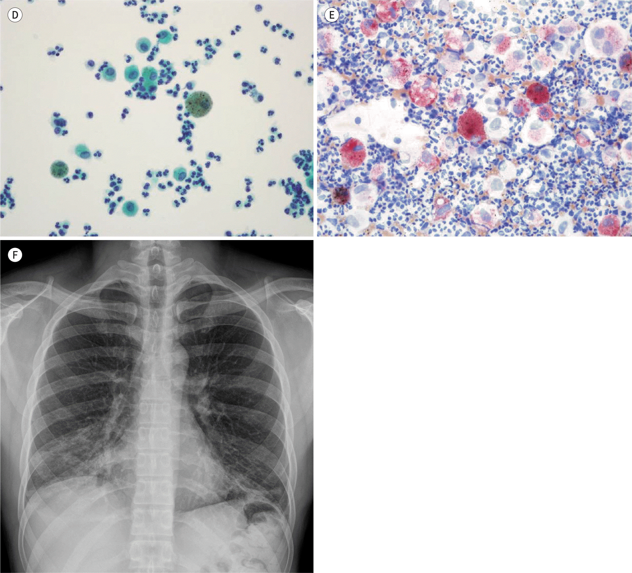
Acute exogenous lipoid pneumonitis is a kind of chemical pneumonitis following the aspiration of volatile hydrocarbon compounds. The clinical and radiological findings are nonspecific. However, it can be diagnosed by the presence of lipid-laden macrophages in bronchoalveolar lavage fluid on the basis of a history of petroleum-based product aspiration. Herein, we report acute exogenous lipoid pneumonitis after unintentional aspiration of diesel fuel during siphon-age in a 31-year-old male. Initially the patient had cough, chest pain, and blood-tinged sputum. The purpose of this case report is to review the radiologic manifestations and the previous literatures.
Go to : 
References
1. Gondouin A, Manzoni P, Ranfaing E, Brun J, Cadranel J, Sadoun D, et al. Exogenous lipid pneumonia: a retrospective multicentre study of 44 cases in France.Eur Respir J. 1996; 9:1463–1469.
2. Marchiori E, Zanetti G, Mano CM, Hochhegger B. Exogenous lipoid pneumonia. Clinical and radiological manifestations.Respir Med. 2011; 105:659–666.
3. Betancourt SL, Martinez-Jimenez S, Rossi SE, Truong MT, Carrillo J, Erasmus JJ. Lipoid pneumonia: spectrum of clinical and radiologic manifestations. AJR Am J Roentgenol. 2010; 194:103–109.

4. Yi MS, Kim KI, Jeong YJ, Park HK, Lee MK. CT findings in hydrocarbon pneumonitis after diesel fuel siphonage. AJR Am J Roentgenol. 2009; 193:1118–1121.

5. Grossi E, Crisanti E, Poletti G, Poletti V. Fire-eater's pneumonitis. Monaldi Arch Chest Dis. 2006; 65:59–61.

6. Franzen D, Kohler M, Degrandi C, Kullak-Ublick GA, Ceschi A. Fire eater's lung: retrospective analysis of 123 cases reported to a National Poison Center.Respiration. 2014; 87:98–104.
7. Gowrinath K, Shanthi V, Sujatha G, Murali Mohan KV. Pneumonitis following diesel fuel siphonage.Respir Med Case Rep. 2012; 5:9–11.
Go to : 
 | Fig. 1.Imaging and histology findings of acute exogenous lipoid pneumonia following diesel fuel siphonage in a 31-year-old man. A. On the day of visit, chest radiograph reveals ill-defined increased opacity in both lower lung zones. B, C. Chest CT scans show multifocal patchy and lobar ground glass opacities and consolidation with interlobular septal thickening in right middle lobe, left upper lobe lingular segment, and both lower |
 | Fig. 1.Imaging and histology findings of acute exogenous lipoid pneumonia following diesel fuel siphonage in a 31-year-old man. D, E. Histopathologic examination of bronchoalveolar lavage fluid. A smear shows many neutrophils and foamy macrophages (oil red O stain, × 400) (D). The cytoplasm is full of red-staining cytoplasmic vacuoles filled with lipid that displaced nucleus to the periphery (oil red O stain, × 400) (E). F. Ten days after the admission, follow-up chest radiograph shows improvement of ill-defined increased opacity in both lower lungs with residual lesions. |


 PDF
PDF ePub
ePub Citation
 Print
Print


 XML Download