

 PDF
PDF ePub
ePub Citation
 Print
Print


Abstract
Purpose
To report a case of iridocorneal endothelial syndrome, which overlapped with some of the features of posterior polymorphous corneal dystrophy.
Case summary
A 61-year-old female presented with tearing pain and blurred vision in her left eye, which was aggravated in the morning. The symptom started approximately 1 year prior to her visit. At the initial visit, the visual acuities were 1.0 in both eyes and the intraocular pressures were normal. On slit-lamp examination, a single pair of horizontal parallel lines was observed at the central corneal endothelial layer in the right eye. In contrast, multiple pairs of oblique parallel lines were observed in the left eye. The lines of the lesions were more prominent and wavier in the left eye than those of the right eye. The overlying cornea was clear, and the corneal thicknesses were in the normal range in both eyes. Using a gonioscopic examination, localized peripheral anterior synechiae were observed only in the left eye. The pupil and iris were normal in both eyes. On specular microscopic examination, the corneal endothelial cell size in the right eye increased and the corneal endothelial density decreased to 668 cells/mm2. In the left eye, multiple abnormal endothelial cells with dark-light reversal were observed. In conclusion, the patient was subsequently diagnosed with iridocorneal syndrome, rather than posterior polymorphous corneal dystrophy.
Figures and Tables
Figure 1
Slit-lamp photography of both eyes at initial examination. (A) In the right eye, a single pair of horizontal parallel lines were observed at corneal endothelial layer (arrows). (B) In the left eye, the multiple pairs of parallel lines were observed at endothelial layer. The lines were running obliquely through the center of the cornea (arrowheads).
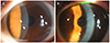
Figure 2
Anterior segment optical coherence tomography of both eyes. The lesions were protruded from the endothelial layer. The lesions were more prominent in the left eye (arrowheads) compared to that of right eye (arrow).
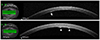
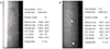
Figure 3
Specular microscopy of both eyes at initial examination. (A) In the right eye, the endothelial cells became large and the density of the cells decreased to 668 cells/mm2. (B) In the left eye, the normal hexagonal shape of the endothelial cells disappeared. The multiple abnormal endothelial cells with dark-light reversal were observed (arrows).
References
1. Feng B, Tang X, Chen H, et al. Unique variations and characteristics of iridocorneal endothelial syndrome in China: a case series of 58 patients. Int Ophthalmol. 2018; 38:2117–2126.

2. Sacchetti M, Mantelli F, Marenco M, et al. Diagnosis and management of iridocorneal endothelial syndrome. Biomed Res Int. 2015; 2015:763093.
3. Lefebvre V, Sowka JW, Frauens BJ. The clinical spectrum between posterior polymorphous dystrophy and iridocorneal endothelial syndromes. Optometry. 2009; 80:431–436.
4. Anderson NJ, Badawi DY, Grossniklaus HE, Stulting RD. Posterior polymorphous membranous dystrophy with overlapping features of iridocorneal endothelial syndrome. Arch Ophthalmol. 2001; 119:624–625.
5. Kim NH, Kim MS. The clinical features and progression of the disease in posterior polymorphous corneal dystrophy (PPCD). J Korean Ophthalmol Soc. 2014; 55:368–373.
6. Grayson M. The nature of hereditary deep polymorphous dystrophy of the cornea: its association with iris and anterior chamber dygenesis. Trans Am Ophthalmol Soc. 1974; 72:516–559.
7. Cibis GW, Tripathi RC. The differential diagnosis of Descemet's tears (Haab's striae) and posterior polymorpous dystrophy bands. A clinicopathologic study. Ophthalmology. 1982; 89:614–620.
8. Yanoff M. Iridocorneal endothelial syndrome: unification of a disease spectrum. Surv Ophthalmol. 1979; 24:1–2.
9. Wilson MC, Shields MB. A comparison of the clinical variations of the iridocorneal endothelial syndrome. Arch Ophthalmol. 1989; 107:1465–1468.
10. Laganowski HC, Kerr Muir MG, Hitchings RA. Glaucoma and the iridocorneal endothelial syndrome. Arch Ophthalmol. 1992; 110:346–350.
11. Chiou AG, Kaufman SC, Beuerman RW, et al. Confocal microscopy in the iridocorneal endothelial syndrome. Br J Ophthalmol. 1999; 83:697–702.
12. Liskova P, Palos M, Hardcastle AJ, Vincent AL. Further genetic and clinical insights of posterior polymorphous corneal dystrophy 3. JAMA Ophthalmol. 2013; 131:1296–1303.
 XML Download