

 PDF
PDF ePub
ePub Citation
 Print
Print


Abstract
Purpose
To report a case of extensive choroidal effusion following the Valsalva maneuver under consecutive general anesthesia.
Case summary
A 41-year-old man who underwent panretinal photocoagulation with proliferative diabetic retinopathy had pars plana vitrectomy and endolaser photocoagulation under general anesthesia due to vitreous hemorrhage. Urology cooperated as the patient had hematuria; the day after the operation, he was transferred to the urology department. Two days after vitrectomy, the patient had an urgent transurethral bladder tumor resection under general anesthesia with suspicion of bladder tumor. At 6 days postoperatively, extensive choroidal effusion was observed from 8 to 10 o'clock on fundus examination and ultrasonography. On day 23 after urological surgery, the choroidal effusion had disappeared without treatment.
Figures and Tables
 | Figure 1Wide-field fundus photography and B-scan ultrasonography of the right eye. (A, B) Choroidal detachment was shown at temporal quadrant (arrowheads) 6 days after vitrectomy (4 days after 2nd general anesthesia). (C, D) Choroidal detachment disappeared without treatment 25 days after vitrectomy (23 days after 2nd general anesthesia).
|
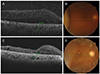 | Figure 2Optical coherence tomography scans showing choroidal thickness of the right eye. Choroidal thickness was 374 µm, (A, B) 6 days after operation (4 days after 2nd general anesthesia). Choroidal thickness decreased by 281 µm (C, D) 25 days after operation (23 days after 2nd general anesthesia). Choroidal effusion disappeared without treatment.
|
References
1. Elagouz M, Stanescu-Segall D, Jackson TL. Uveal effusion syndrome. Surv Ophthalmol. 2010; 55:134–145.

2. Schepens CL, Brockhurst RJ. Uveal effusion. I. Clinical picture. Arch Ophthalmol. 1963; 70:189–201.
3. Gass JD, Jallow S. Idiopathic serous detachment of the choroid, ciliary body, and retina (uveal effusion syndrome). Ophthalmology. 1982; 89:1018–1032.
4. Gass JD. Uveal effusion syndrome: a new hypothesis concerning pathogenesis and technique of surgical treatment. Trans Am Ophthalmol Soc. 1983; 81:246–260.
5. Kumar A, Kedar S, Singh RP. The indocyanine green findings in idiopathic uveal effusion syndrome. Indian J Ophthalmol. 2002; 50:217–219.
6. Jones WL. Valsalva maneuver induced vitreous hemorrhage. J Am Optom Assoc. 1995; 66:301–304.
7. Lim HW, Ko BW, Song YM, Lee BR. Suprachoroidal hemorrhage during pars plana vitrectomy associated with valsalva maneuver. J Korean Ophthalmol Soc. 2008; 49:1022–1027.
8. Jung EY, Kim IJ, Lee EC. A case of recurrent valsalva retinopathy associated with exercising a barbell. J Korean Ophthalmol Soc. 2004; 45:1040–1044.
 XML Download