

 PDF
PDF ePub
ePub Citation
 Print
Print


Abstract
Purpose
To evaluate the effect of epiretinal membrane (ERM) on the outcomes of intravitreal dexamethasone implant (Ozurdex®, Allergan, Irvine, CA, USA) treatment for macular edema (ME) secondary to branch retinal vein occlusion (BRVO).
Methods
Thirty eyes of 30 patients who received Ozurdex treatment for ME secondary to BRVO, and were followed-up for at least 6 months were retrospectively reviewed. Patients were divided into two groups based on the presence (ERM [+] or absence ERM [−]) of ERM at baseline. The best-corrected visual acuity (BCVA), central foveal thickness (CFT), recurrence of ME, and retreatment rate were evaluated at baseline, 1, 3, and 6 months after Ozurdex injection.
Results
Ten eyes of 30 eyes (33%) showed ERM at baseline. While the mean CFT was significantly reduced at 1 month after Ozurdex injection, it began to increase gradually thereafter in both groups. The ERM (+) group showed a significantly higher mean CFT than the corresponding values of the ERM (−) group at 1 (p = 0.022) and 6 months (p = 0.001) after Ozurdex injection. However, no significant difference was found in the BCVA between the two groups at every visit. The proportion of eyes with ME was significantly higher in the ERM (+) group (90%) than that in the ERM (−) group (35%) at 6 months after Ozurdex injection (p = 0.009). There were no significant differences between the two groups in the percentage of retreatment, time to retreatment, and type of materials used for retreatment.
Figures and Tables
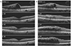 | Figure 1Representative optical coherence tomography images in patients with branch retinal vein occlusion associated with macular edema and treated with intravitreal dexamethasone implant (Ozurdex®, Allergan, Irvine, CA, USA). (A) The eye without epiretinal membrane (ERM) showed severe macular edema at baseline. After Ozurdex injection, no significant recurrence was observed during the 6-month of follow-up period. (B) The eye with ERM also showed significant macular edema at baseline. The macular edema absorbed at 1 month after Ozurdex injection but recurred at 3 months. At 3 months, an additional injection of bevacizumab was performed, but the macular edema persisted at 6 months.
|
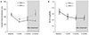 | Figure 2Chronological changes in mean central foveal thickness (CFT) (A) and best-corrected visual acuity (BCVA) (B) in ERM (−) and ERM (+) groups. (A) The ERM (+) group showed significantly higher mean CFT at 1 and 6 months after Ozurdex injection than the corresponding values of ERM (−) group. (B) There were no significant differences in BCVA between the two groups at every visit. *p<0.05, **p<0.01 for comparisons between groups at each follow-up visit.
|
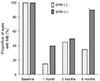 | Figure 3The proportion of eyes with macular edema in ERM (−) and ERM (+) groups. It was significantly higher in ERM (+) group than that of ERM (−) group at 6 months after Ozurdex injection. ME = macular edema. *p < 0.01 for comparisons between groups at each follow-up visit.
|
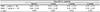
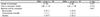
Table 1
Demographics and baseline characteristics of included patients

Values are presented as mean ± standard deviation or number.
ERM (−) group = eyes without epiretinal membrane; ERM (+) group = eyes with epiretinal membrane; CFT = central foveal thickness; BCVA = best-corrected visual acuity; logMAR = logarithm of the minimal angle of resolution.
*Pearson's chi-squared test, †Mann-Whitney U-test.
![]()

References
1. Orth DH, Patz A. Retinal branch vein occlusion. Surv Ophthalmol. 1978; 22:357–376.

2. Zhao J, Sastry SM, Sperduto RD, et al. Arteriovenous crossing patterns in branch retinal vein occlusion. The Eye Disease Case-Control Study Group. Ophthalmology. 1993; 100:423–428.
3. Weinberg D, Dodwell DG, Fern SA. Anatomy of arteriovenous crossings in branch retinal vein occlusion. Am J Ophthalmol. 1990; 109:298–302.
4. The Branch Vein Occlusion Study Group. Argon laser photocoagulation for macular edema in branch vein occlusion. Am J Ophthalmol. 1984; 98:271–282.
5. Pe'er J, Folberg R, Itin A, et al. Vascular endothelial growth factor upregulation in human central retinal vein occlusion. Ophthalmology. 1998; 105:412–416.
6. Coscas G, Loewenstein A, Augustin A, et al. Management of retinal vein occlusion--consensus document. Ophthalmologica. 2011; 226:4–28.
7. Glacet-Bernard A, Coscas G, Zourdani A, et al. Steroids and macular edema from retinal vein occlusion. Eur J Ophthalmol. 2011; 21 Suppl 6:S37–S44.
8. Haller JA, Bandello F, Belfort R Jr, et al. Randomized, sham-controlled trial of dexamethasone intravitreal implant in patients with macular edema due to retinal vein occlusion. Ophthalmology. 2010; 117:1134–1146.e3.
9. Chatziralli I, Stavrakas P, Theodossiadis G, et al. The impact of epiretinal membrane in neovascular age-related macular degeneration treatment: a spectral-domain optical coherence tomography study. Semin Ophthalmol. 2018; 33:651–656.
10. Kulikov AN, Sosnovskii SV, Berezin RD, et al. Vitreoretinal interface abnormalities in diabetic macular edema and effectiveness of anti-VEGF therapy: an optical coherence tomography study. Clin Ophthalmol. 2017; 11:1995–2002.
11. Hayreh SS, Zimmerman MB. Fundus changes in branch retinal vein occlusion. Retina. 2015; 35:1016–1027.
12. Mitchell P, Smith W, Chey T, et al. Prevalence and associations of epiretinal membranes. The Blue Mountains Eye Study, Australia. Ophthalmology. 1997; 104:1033–1040.
13. Marticorena J, Romano MR, Heimann H, et al. Intravitreal bevacizumab for retinal vein occlusion and early growth of epiretinal membrane: a possible secondary effect? Br J Ophthalmol. 2011; 95:391–395.
14. Stevenson W, Prospero Ponce CM, Agarwal DR, et al. Epiretinal membrane: optical coherence tomography-based diagnosis and classification. Clin Ophthalmol. 2016; 10:527–534.
15. Sugar EA, Jabs DA, Altaweel MM, et al. Identifying a clinically meaningful threshold for change in uveitic macular edema evaluated by optical coherence tomography. Am J Ophthalmol. 2011; 152:1044–1052.e5.
16. Gass J. Macular dysfunction caused by epiretinal membrane contraction. In : Gass JDM, editor. Stereoscopic Atlas of Macular Diseases: Diagnosis and Treatment. 4th ed. St. Louis: Mosby;1997. p. 938–951. v. 2.
17. Boyer DS, Yoon YH, Belfort R Jr, et al. Three-year, randomized, sham-controlled trial of dexamethasone intravitreal implant in patients with diabetic macular edema. Ophthalmology. 2014; 121:1904–1914.
18. Myung JS, Aaker GD, Kiss S. Treatment of noninfectious posterior uveitis with dexamethasone intravitreal implant. Clin Ophthalmol. 2010; 4:1423–1426.
19. Zhao F, Gandorfer A, Haritoglou C, et al. Epiretinal cell proliferation in macular pucker and vitreomacular traction syndrome: analysis of flat-mounted internal limiting membrane specimens. Retina. 2013; 33:77–88.
20. Cho HJ, Kim JM, Kim HS, et al. Effect of epiretinal membranes on antivascular endothelial growth factor treatment for neovascular age-related macular degeneration. J Ocul Pharmacol Ther. 2017; 33:452–458.
21. Wong Y, Steel DHW, Habib MS, et al. Vitreoretinal interface abnormalities in patients treatedwith ranibizumab for diabetic macular oedema. Graefes Arch Clin Exp Ophthalmol. 2017; 255:733–742.
22. Lee SJ, Koh HJ. Effects of vitreomacular adhesion on anti-vascular endothelial growth factor treatment for exudative age-related macular degeneration. Ophthalmology. 2011; 118:101–110.
23. Nomura Y, Takahashi H, Tan X, et al. Effects of vitreomacular adhesion on ranibizumab treatment in Japanese patients with age-related macular degeneration. Jpn J Ophthalmol. 2014; 58:443–447.
 XML Download