

 PDF
PDF ePub
ePub Citation
 Print
Print


INTRODUCTION
Various morphologic features of calcifications are one of the important criteria determining malignancy of breast tumor according to the American College of Radiology Breast Imaging Reporting and Data System (ACR-BIRADS) 5th edition (1). The interpretation of these calcifications, however, is partly dependent on the descriptors, and different diagnostic confirmation can be demonstrated. A mass with almost exclusive portion of calcification can be easily considered as a dense benign calcification. In the ACR-BIRADS, classic and large (> 2–3 mm in greatest diameter) coarse or “popcorn-like” calcification is defined as category 2 benign finding, and the recommended management is only regular follow-up. Subsequently, it can be more extensive and confluent and when so densely calcified, the underlying mass usually is not visible (1). However, there have been a few reported cases of primary and secondary breast cancer presenting as a mass replaced by calcification, which could be one of the causes of misdiagnosis on mammography. We present two cases of a mass replaced by calcification that resulted in malignant breast tumor. The imaging features of mammography, ultrasound (US), and magnetic resonance imaging (MRI) are presented and correlated pathologically.
CASE REPORT
CASE 1
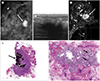
A 50-year-old woman presented to our breast center for evaluation of a palpable lump in the upper inner portion of right breast that was increasing in size for a year. Right mammogram showed extremely dense composition and a 24 mm-sized, calcified mass with an oval shape and circumscribed margins in the clinically palpable upper inner area. Coarse calcification was also noted in the upper outer portion (Fig. 1A). The mammographic BI-RADS assessment was category 2, but additional breast US was performed because of the growing palpable breast lump. US revealed a 26 mm-sized, irregular, and markedly hypoechoic mass with indistinct margins and strong posterior shadowing in the palpable area, which was a suspicious finding for malignancy (Fig. 1B). The BI-RADS assessment between the mammography and US findings was discordant, and thus US-guided 14-gauge core needle biopsy was performed. The pathologic result was consistent with invasive micropapillary carcinoma. The subtraction image of dynamic contrast-enhanced breast MRI showed an oval shaped mass with circumscribed margins and heterogeneous enhancement and a satellite enhancing nodule. The kinetic curve showed initial fast and delayed plateau enhancement pattern (Fig. 1C). The mass had a portion with low signal intensity on both T1 and T2-weighted image, suggesting calcification (not shown). She underwent total mastectomy, and the gross specimen measured 2.2 × 1.8 cm and showed a grayish granular appearance with a myxoid cut surface. Microscopically, it was diagnosed as mixed carcinoma that was composed of mucinous carcinoma (60%) with micropapillary carcinoma (40%) (Fig. 1D, E). Calcifications were noted in both cancers.
CASE 2
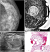
A 46-year-old woman visited an another clinic for screening, and the mammogram had shown heterogeneously dense composition and a 13 mm-sized mass almost entirely substituted by calcification with circumscribed margins in the left central area (Fig. 2A). Considered that no findings suspicious of malignancy were noted, she was recommended only for regular follow-up. After 2 years, she visited the clinic because of a palpable lump in the left breast. Left magnification mammogram showed that the pre-existing mass increased in size to 25 mm; new grouped calcifications in the subareolar area were also noted (Fig. 2B). Breast US showed that the mass was an oval-shaped hyperechoic mass with a few internal echogenic calcifications and circumscribed margins (Fig. 2C). Pathological findings of excisional biopsy indicated mucinous carcinoma, measuring 2.0 × 1.5 cm (Fig. 2D). She was referred to our breast center for operative treatment and underwent breast conservation surgery. There was no residual cancer, and axillary sentinel node biopsy was negative. This was an interval breast cancer that had been assessed as category 2 benign finding at the first screening mammography.
DISCUSSION
Some cases of primary and secondary malignant breast tumor that appeared as a mass mimicking benign finding with extensive portion of calcification on mammography have been reported. To our knowledge, there have been cases of metaplastic breast carcinoma, mucinous carcinoma, primary and secondary osteosarcoma, and metastasis from ovarian cancer.
Metaplastic carcinoma of the breast is rare, accounting for only 5% of all breast carcinomas. It shows metaplastic change in the form of mesenchymal elements producing osseous matrix and has squamous cell and spindle cell components (2). Meanwhile, osseous or chondroid metaplasia can appear as a densely calcified mass. Lee et al. (3) reported a case of metaplastic breast carcinoma with osseous differentiation that as appeared as a largely calcified mass with a partially spiculated margin. Although it could have been mistaken for as a benign mass because of the dense calcification, it could be assessed as suspicion for malignancy due to the spiculated margin. The dense calcification was confirmed as ossifying osteoid matrix. Another case reported by Evans et al. (4) showed a densely calcified mass with a spongy, bonelike appearance along the periphery of the calcification on mammogram. An additional image obtained using a higher kilovoltage setting showed trabeculations, which are consistent with an osteoid matrix.
Mucinous carcinoma is even rarer and accounts for only approximately 1.5–2.0% of all breast carcinoma (5). It has two subtypes according to histologic features: the pure type containing typical extracellular mucin-producing cells mostly and the mixed type with much lower portion of this cells (50–90%) (35). The typical imaging finding is a well-circumscribed round or oval-shaped mass on mammography and rarely shows calcification. When present, the calcification shows pleomorphic and clustered form, and they also appear clumped and amorphous (6). In the current report, both cases of mixed- and pure-type mucinous carcinoma presented as a more exclusively calcified mass than the usual calcification patterns reported previously. Although there was an atypical case of breast mucinous carcinoma with coarse calcification (6), it has not been reported to have almost entirely calcified portion of the mass. As a mass replaced by calcification or a mass accompanying benign coarse calcification is generally considered as benign lesion, the malignant breast tumor with these characteristics are prone to be a missed cancer. The first case of this report was also assessed as category 2 benign finding on mammography. However, she had a palpable breast lump, and subsequent breast US showed suspicious features for malignancy. The second case was an interval cancer, as shown above, because of the circumscribed marginated mass replaced by calcification on the first screening mammography.
Primary osteosarcoma of the breast is extremely rare and usually reveals a heavily calcified mass as a result of intermembranous ossification with irregular margin (7). Metastatic osteosarcoma to the breast was also described to have dense calcification in a case reported by Kim et al., (8) even though metastatic osteosarcoma usually involves lung and skeleton.
Calcifications in metastatic tumor of the breast are uncommon, except in ovarian metastasis. Metastatic lesion in the breast represents clusters of smooth and irregular dense calcification from the ovarian tumor. They are suggested to be related with psammoma bodies of malignant tumor (9).
In conclusion, a mass replaced by calcification on mammography can be easily considered as benign finding, however, we should keep in mind the possibility of malignancy such as mucinous breast cancer and assess the mass thoroughly with other suspicious findings for malignancy such as partially non-circumscribed margins or clinical palpability to prevent missed cancer.
 XML Download