Abstract
High intensity focused ultrasound (HIFU) is a non-surgical and non-invasive treatment option in patients with uterine myoma and adenomyosis. As the use of HIFU increases in the clinical practice, it is important to be aware of imaging findings related to ultrasound (US)-guided HIFU ablation and its potential complications. However, there are few reports on the imaging findings regarding complications of US-guided HIFU ablation. Here, we report a case of acute complication after US-guided HIFU ablation, surgically confirmed as thermal injury with necrosis of skin, subcutaneous tissue, anterior abdominal wall muscles, peritoneum and uterus.
High-intensity focused ultrasound (HIFU) ablation is performed using ultrasound (US) waves to transfer energy through tissue (1). Currently, US-guided HIFU ablation is regarded as a non-surgical and non-invasive treatment option for fibroids and adenomyosis in women who want to preserve fertility (2). Although safe and effective, US-guided HIFU ablation is known to cause several mild and severe complications (134). Despite this, there are few reports on the imaging findings of complications following US-guided HIFU ablation. We present CT findings of acute complications associated with US-guided HIFU ablation in a patient who visited the emergency department with abdominal pain.
A 44-year-old female patient presented to the emergency department with diffuse low abdominal pain and fever. The patient had undergone ablation of the uterine adenomyosis using a US-guided HIFU device (YDME FEP-BY02; Yuande Bio-Medical Engineering, Beijing, China) at the outside private hospital three days prior to the presentation. On physical examination, there were rebound tenderness on palpation and visible skin burn. Speculum examination showed pus-like discharge through the cervix. The patient underwent contrast enhanced abdominal CT to find out if there are any US-guided HIFU ablation-related injuries in the abdominal and pelvic organs.
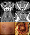
The CT showed the sharply demarcated zone of decreased density with linear lateral margins at the anterior portion of the uterus (Fig. 1A–D). In addition, there were hypodense areas with well-demarcated edges, which extend to bilateral rectus abdominal muscles undeniably reflecting the characteristic ablation field used in US-guided HIFU ablation. The CT also showed overlying skin thickening, non-enhancing thickening of the superficial and deep fascia at the anterior pelvic wall, peritoneal thickening with fat infiltration, and small amount of extraperitoneal fluid collection in the pelvic cavity. However, the urinary bladder and bowel that were considered within the ablation field were not affected. These findings suggest a thermal injury with necrosis of the skin, subcutaneous tissue, anterior abdominal wall muscles, peritoneum, and uterus.
The patient underwent an emergency operation. On inspection, there were skin necrosis and dark brownish lesions on the anterior wall of the uterus (8 × 8 cm) presumably due to thermal injury (Fig. 1E, F). The rectus muscle and deep fascia were also non-viable. The patient underwent total abdominal hysterectomy and debridements of muscle necrosis and skin burn, followed by CuraVAC (Daewoong Pharmaceutical, Co., Ltd., Seoul, Korea) application and skin closure. Hemorrhage and necrosis in endometrium and myometrium were pathologically confirmed. The patient was discharged without postoperative complications. Subsequently, the skin burn improved with continuous outpatient clinic visits at the department of the plastic surgery.
The principle of HIFU therapy is to center the US energy on the target, causing heating, cavitation, and direct damage to tumor blood vessels, resulting in tissue ablation. Therefore, high-energy US waves passing through the focal therapeutic zone can induce damages to internal organs just anterior or posterior to the focal zone and overlying soft tissue including the skin. However, the majority of complications occurring after US-guided HIFU ablation are caused by reflections of US waves due to (bowel) gas or bone structures. Skin burns can be caused by poor acoustic windows between the skin and the therapeutic window or by previous operation scars. Other possible causes include inaccurate targeting, excessive power generation, mechanical causes, or unskilled operator. In the monitoring of therapy, unlike magnetic resonance (MR)-guided HIFU ablation which quantifies changes in temperature and thermal dose of the treated tissue directly, US-guided HIFU ablation evaluates adequacy of ablation based on the changes in grayscale or echogenicity (12). However, changes in sonographic echogenicity are not a direct measure of temperature elevation and merely reflect acoustic cavitation or tissue boiling. Thus, there may be a mismatch between the region where the actual coagulation necrosis occurs and the region where the echogenicity changes (1). The sonographic monitoring during ablation has the drawbacks of relatively poor tissuecontrast, limited field of view and progressive deterioration of image quality as the treatment continues. Therefore, the difficulty in targeting the organs, the drawbacks of therapy monitoring and poor sonic window by bone or bowel gas can potentially increase the risk of side effects of US-guided HIFU ablation.
Previous studies classified complications of US-guided HIFU ablation into mild complications that can be conservatively managed and severe adverse events that need surgical repair with subsequent sequelae. The incidence and types of side effects of US-guided HIFU-ablation vary based on previous studies. Five of the 11 studies reported no adverse events or serious complications (2). Chen et al. (4) reported outcome of the total 9988 patients receiving US-guided HIFU ablation, of which 1273 (12.75%) required no treatment or observation only, 26 (0.26%) required a minor hospitalization, and only 6 (0.06%) required major therapy. Severe events requiring major therapy were acute renal failure, intestinal perforation, and hernia in the abdominal wall. Zhang et al. (5) reported other severe adverse events such as skin burn requiring surgical repair, bladder injury, and deep vein thrombosis. In addition to the acute complication of HIFU ablation, delayed complications may also occur. Kim et al. (6) reported delayed complications such as rapid uterine enlargement and heavy vaginal bleeding 8 months following HIFU treatment. Quinn et al. (7) reported that 59.3% of patients required additional treatments (hysterectomy, myomectomy, and uterine artery embolization) within 5 years after HIFU therapy. Ko et al. (8) reported a case of thermal injury to the bowel which resulted in perforation after US-guided HIFU ablation for uterine adenomyosis. However, there have been few reports illustrating the image finding of these complications. Most reports of imaging findings relating to US-guided HIFU ablation have focused on the post-procedural evaluation of the ablation zone after the procedure using contrast enhanced sonography or contrast-enhanced MR (910).
Our case demonstrates the CT image findings of acute, severe thermal injury of abdominal wall and uterus after US-guided HIFU ablation for uterine adenomyosis. Hwang et al. (3) reported imaging findings of delayed intestinal perforation and necrosis of the rectus muscle 29 days after HIFU treatment for uterine leiomyoma. The CT image findings of our case were similar to Hwang's report except bowel perforation in that there were hypodense areas with linear well-demarcated edges which are thought to correspond to the margin of the characteristic ablation field used in US-guided HIFU ablation. However, unlike Hwang's report, our report presents a rare case of severe thermal injury in its acute phase after US-guided HIFU ablation. The complications of HIFU ablation are very rare and most cases are with minor gynecologic symptoms. However, regardless of when the procedure was performed, clinicians should consider the possibility of serious complication related to US-guided HIFU ablation in patients who complain of gynecologic symptom and abdominal pain with previous history of US-guided HIFU ablation. Radiologists should understand the mechanism of complications of US-guided HIFU therapy (i.e., the organs injury within ablation field due to excessive power generation or inaccurate targeting and complications by the reflection of US waves due to bowel gas or bone structures within the narrow pelvic cavity) and carefully review images of organs such as skin, small bowel, uterus, vascular structure, and urinary bladder that can be potentially damaged.
Most of the previous reports have focused on the types and incidence of complications of US-guided HIFU ablation without CT findings or treatment monitoring. In this case, we present CT findings of acute complications associated with US-guided HIFU ablation in a patient who presented to the emergency department with abdominal pain. We review the mechanism of complications of US-guided HIFU therapy in order to alert the radiologists to pay close attention to the possible serious complications of US-guided HIFU ablation. The radiologists should carefully evaluate structures within focal zone of US-guided HIFU ablation and also consider the injuries to adjacent organs by the reflection of US waves.
Figures and Tables
 | Fig. 1Complication following ultrasound-guided HIFU ablation for uterine adenomyosis in a 44-year-old woman, presenting with low abdominal pain.
A, B. Axial CT image shows overlying skin thickening, non-enhancing thickening of the superficial and deep fascia at the anterior pelvic wall, peritoneal thickening with fat infiltration in the pelvic cavity, and non-enhancement at bilateral rectus abdominis muscles (A, B; arrowheads). Note the sharply demarcated zone of hypodensity with linear lateral margins at the anterior portion of the uterus (B, arrow).
C, D. Coronal reconstructed CT image shows area of non-enhancement at the bilateral rectus abdominis muscles (C, arrowheads) and zone of hypodensity at the anterior portion of the uterus (D, arrow).
E, F. Photos show (E) skin necrosis in the treated area and (F) thermal injury with necrosis (arrow) on the uterine anterior wall in the surgical field.
HIFU = high intensity focused ultrasound
|
References
1. Kim YS, Rhim H, Choi MJ, Lim HK, Choi D. High-intensity focused ultrasound therapy: an overview for radiologists. Korean J Radiol. 2008; 9:291–302.

2. Cheung VY. Current status of high-intensity focused ultrasound for the management of uterine adenomyosis. Ultrasonography. 2017; 36:95–102.

3. Hwang DW, Song HS, Kim HS, Chun KC, Koh JW, Kim YA. Delayed intestinal perforation and vertebral osteomyelitis after high-intensity focused ultrasound treatment for uterine leiomyoma. Obstet Gynecol Sci. 2017; 60:490–493.

4. Chen J, Chen W, Zhang L, Li K, Peng S, He M, et al. Safety of ultrasound-guided ultrasound ablation for uterine fibroids and adenomyosis: a review of 9988 cases. Ultrason Sonochem. 2015; 27:671–676.

5. Zhang L, Zhang W, Orsi F, Chen W, Wang Z. Ultrasound-guided high intensity focused ultrasound for the treatment of gynaecological diseases: a review of safety and efficacy. Int J Hyperthermia. 2015; 31:280–284.

6. Kim HK, Kim D, Lee MK, Lee CR, Kang SY, Chung YJ, et al. Three cases of complications after high-intensity focused ultrasound treatment in unmarried women. Obstet Gynecol Sci. 2015; 58:542–546.

7. Quinn SD, Vedelago J, Gedroyc W, Regan L. Safety and five-year re-intervention following magnetic resonance-guided focused ultrasound (MRgFUS) for uterine fibroids. Eur J Obstet Gynecol Reprod Biol. 2014; 182:247–251.

8. Ko JK, Seto MT, Cheung VY. Thermal bowel injury after ultrasound-guided high-intensity focused ultrasound for uterine adenomyosis. Ultrasound Obstet Gynecol. 2018; 52:282–283.
9. Cheng CQ, Zhang , RT , Xiong Y, Chen L, Wang J, Huang GH, et al. Contrast-enhanced ultrasound for evaluation of high-intensity focused ultrasound treatment of benign uterine diseases: retrospective analysis of contrast safety. Medicine (Baltimore). 2015; 94:e729.


 PDF
PDF ePub
ePub Citation
 Print
Print


 XML Download