

 PDF
PDF ePub
ePub Citation
 Print
Print


INTRODUCTION
Extraskeletal Ewing's sarcoma (EES), especially involving spine accounts for only 5% of all ES (1). It is known to be found mainly in the paravertebral and epidural space (2). And ES usually occurs in children or young adults (1). Primary EES of spinal nerve has been reported only a few cases and there has been only one case of a senior patient in eighth decade (3).
Herein, we report a case of spinal epidural EES in a 73-year-old female.
CASE REPORT
A 73-year-old female visited to our hospital complaining pain in the posterior neck and left arm. The symptoms started 6 months ago and aggravated 2 days ago. Physical examination revealed hypoesthesia on her left ulnar nerve territory without decrease of muscle strength. She underwent plain radiography and CT of cervical spine. Plain radiographs of the cervical spine showed no significant abnormality other than disc space narrowing in C5–C6 level (not shown).
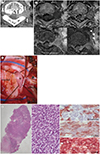
CT revealed a dumbbell-shaped mass in the left neural foramen of T1–T2. No calcification was seen in the mass. There was mild widening of left neural foramen (Fig. 1A). For further evaluation, MRI of the cervical and thoracic spine was performed. MR images showed that the medial part of the mass is in the intradural and extramedullary region and the lateral part of the mass extends to the paravertebral area through the left neural foramen. The lesion shows intermediate signal intensity on T1-weight images and homogenously slightly high signal intensity on T2-weighted images (Fig. 1B). After administration of intravenous contrast material, the mass is enhanced homogeneously. Size of the lesion is measured approximately 2.6 × 1.3 × 1.5 cm. The mass is branching outside the left neural foramen and extending along the first branch of left T1 nerve (Fig. 1B).
Based on the CT and MRI findings, the benign nerve sheath tumor was suggested. And then, the patient underwent surgery. In the operation, the tumor was revealed after laminectomy of T1 vertebra and also after incision of dura (Fig. 1C). The mass was strongly attached to the nerve root, partly intermingled with the nerve fibers. Subtotal resection of the tumor was done with moter evoked potential monitoring to minimize injury of the nerve.
Microscopic examination revealed that the tumor was composed of alternating highly cellular and paucicellular components with dense collagen bands (× 10). The high-power field view showed highly overlapping blue round cells with oval nuclei and scanty cytoplasm, compatible with blue round cell tumor. No evidence of neuroectodermal differentiation, such as rosette formation was identified (× 400) (Fig. 1D). The result of immunohistochemical staining revealed weak positivity for vimentin and CD56, and strong membranous staining for CD99, compatible with ES (Fig. 1D). Further gene mutation study revealed EWSR1 gene translocation and the final pathological diagnosis was made as EES.
After confirmative diagnosis, the patient transferred to cancer clinic for adjuvant chemotherapy and radiation therapy. After twelve months from the surgery, the tumor was reduced in size and the patient was in stable condition without local recurrence, only complaining mild tingling sensation on her elbow. However, recently, after post-operative sixteen months, local recurrence and leptomeningeal seeding developed and the patient is receiving chemotherapy.
DISCUSSION
EES is a rare form of round cell malignant tumor with extraskeletal origin (124). It accounts for about 10% of ES in children and 5% of ES in adults (3). The primary site of EES involving extradural and intradural extramedullary space is extremely rare (12345678910). Prefered anatomic origin of EES is paravertebral and epidural space, so associated symptoms are variable such as local pain, gait disturbance, motor deficit, radicular pain, etc (145).
Imaging features of EES are nonspecific but, usually appears solid, iso signal intensity on T1-weighted image, intermediate to high signal intensity on T2-weighted image and moderate to strong enhancement after gadolinium administration (12345678910). Some reported lesions show slightly inhomogeneous signal intensity or low signal intensity on T2-weighted images (1345) and heterogeneous enhancement (1356).
In our case, the mass was iso signal intensity in T1-weighted image and slightly high signal intensity in T2-weighted image with marked enhancement, similar to previous cases.
The radiologic differential diagnosis of EES involving epidural space includes benign nerve sheath tumor such as schwannoma, lymphoma and malignant nerve sheath tumor (2347). Schwannoma has smooth margin and dumbell shaped with intense enhancement. On T2-weighted image, schwannoma appears heterogeneously high signal intenstiy with target sign (347). Lymphoma involving epidural space originates from body of vertebra or paravertebral lymph node then extends to epidural space. It is slightly high signal intensity to muscle on T1-weighted image and T2-weighted image involving multi-segment of spine with homogeneous enhancement along epidural infiltration (9). Malignant nerve sheath tumor of spine appears expansile soft tissue tumor destructing adjacent vertebra with paraspinal and epidural extension. On MR, the mass appears low signal intensity on T1-weighted image and high signal intensity on T2-weighted image. It is mostly associated with neurofibromatosis type 1 (10).
This case was initially presumed to be a benign nerve sheath tumor, however the final diagnosis was an EES. There was no definite aggressive feature such as ill-defined margin, adjacent bony destruction, heterogeneous signal intensity or attenuation of the mass, or large size of the mass. However, in the literature, there are some reported cases of EES mimicking benign nerve sheath tumors (467) and it was difficult to differentiate EES from benign nerve sheath tumor only with radiologic imaging, without pathologic confirmation in those cases. In our case, the lesion extended to the 1st branch of thoracic nerve root and this feature may indicate relatively more aggressive kinds of tumor such as lymphoma or malignant peripheral nerve sheath tumor rather than benign nerve sheath tumor, even though ES is very rare in elder patients. However, this finding may be seen in neurofibromas.
Therefore, even there is well-marginated dumbbell-shaped extradural mass whick looks benign, if there is subtle ill-defined enhancing or high signal intensity lesion extended to peripheral nerve, possibility of malignancy might be considered.
We reported a case of EES arising from thoracic nerve root in 73-year-old woman. Because of its rarity and varying morphology on imaging modality, it is difficult to diagnosis from radiologic finding before pathologic confirmation. We suggest that in case of nerve root tumor showing atypical radiologic findings of nerve sheath tumors, EES also could be considered as differential diagnoses although these tumors are very rare in elder patients.
 XML Download