

 PDF
PDF ePub
ePub Citation
 Print
Print


INTRODUCTION
Transcatheter arterial chemoembolization (TACE) is widely accepted as an effective procedure for hepatocellular carcinoma (HCC) (1). However, unexpected dissection of the celiac axis and its branches may occur during selective TACE and become an obstruction to the success of the procedure (2). The previously published report has demonstrated that the incidence of iatrogenic dissection during TACE was approximately 0.12% (2).
We encountered HCC necrosis caused by iatrogenic hepatic artery (HA) dissection during TACE. There are no reports describing the HCC necrosis caused by iatrogenic HA dissection in previously published literatures.
CASE REPORT
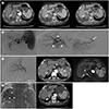
A 67-year-old female, a chronic hepatitis B virus carrier, had a 16-year history of liver cirrhosis. She also had a 7-year history of esophageal varix. She underwent surgical operations for colon cancer 15 years ago and it had been completely cured. On 21 August 2015, she came to our emergency room for hematemesis. Since then, she was followed up for liver cirrhosis and esophageal varix at our hospital. Incidentally, 3-phase abdomen CT for follow up showed a 3.0 × 2.5 cm new nodular lesion with subtle centripetal arterial enhancement and portal/delayed wash-out between Couinaud's segment VI (S6) and VII (S7), compatible with HCC (Fig. 1A). Patient was admitted to our institute to treat the lesion. The 3-phase abdomen CT also showed portal vein thrombosis in right portal vein, main portal vein, splenic vein and proximal superior mesenteric vein. The tumor was located closely in the inferior vena cava. Radio-frequency ablation and surgical resection were not considered due to portal vein thrombosis and tumor location. Lastly, she referred to our department for TACE. Laboratory test results at hospital admission showed levels of aspartate aminotransferase (AST) and alanine aminotransferase (ALT) at 43 U/L and 23 U/L (AST: normal range < 40 U/L, ALT: normal range < 40 U/L), respectively. Total bilirubin (TB) was 1.29 mg/dL (TB: normal range < 1.30 mg/dL) and albumin was 3.70 g/dL (normal range: 3.5–5.2 g/dL). At that time, she had slight ascites but no encephalopathy. The patient's hepatic function was given a Child-Pugh score of 5 points. On laboratory test, alpha-fetoprotein (AFP) level was 2.7 ng/mL (AFP: normal range < 7 ng/mL). The test for protein induced by vitamin K absence or antagonist II (PIVKA-II) was not performed.
During the procedure, proper hepatic arterial angiography showed an arterial staining target lesion in right hepatic lobe (Fig. 1B). Using microguidewire (Transend, Boston Scientific, Natick, MA, USA) and microcatheter (Progreat Alpha, Terumo, Tokyo, Japan), posterior divisional HA angiography with contrast-enhanced cone-beam CT (INFX-8000V, Toshiba, Tokyo, Japan) was performed, and an arterial staining target lesion supplied from subbranch of hepatic arterial branch for S7 (A7) was detected. We tried to advance microcatheter into A7 for selective TACE in vain due to HA dissection of A7 orifice. Angiography did not show arterial flow to S7 including target lesion due to A7 dissection (Fig. 1B). Considering the possibility of flow to the target lesion via hepatic arterial branch for S6 (A6), we tried angiography via A6, but it showed no arterial staining target lesion in S6 (Fig. 1B). Even after the nitroglycerin 10 mg (Nitro, Myungmoon Pharm., Seoul, Korea) injection, no visible arterial flow was shown at A7 on angiography. We interrupted TACE procedure and decided to redo it three weeks later based on our knowledge gained from the past experience that dissected HA recanalized within three weeks.
After one month, posterior divisional HA angiography showed recanalization of previously dissected A7, but no tumor staining was found (Fig. 1C). The TACE procedure was suspended and the patient immediately underwent 3-phase abdomen CT in CT suite. Noncontrast CT showed low-density S7 mass with thin and high-density parts along the rim of the mass. However, there was no enhancement of the mass on 3-phase abdomen CT, which was compatible with HCC necrosis. In addition, the size of HCC decreased (2.5 × 2.2 cm) (Fig. 1C), as well. We concluded that there was no viable HCC and ended the procedure without chemoembolization. In the formal report read by abdominal radiologist a few days later, the target lesion was judged to be HCC necrosis.
After two months, follow-up liver MRI showed central HCC necrosis with peripheral viable tumor in S7 (Fig. 1C). Laboratory testing for follow-up showed levels of AFP and PIVKA-II at 2.9 ng/mL and 12 mAU/mL (PIVKA-II: normal range < 40 mAU/mL), respectively. After the patient referred to our department for TACE, posterior divisional HA angiography showed no staining lesion around the expected target. Nevertheless, we planned to perform blind TACE via A7 with wedging the tip of microcatheter at the orifice of A7 according to MRI finding. Taking previous HA dissection into consideration, selective angiography via A7 was tried carefully for proximal portion of A7. After the angiography, segmentally selective TACE was done for recurred HCC in S7 by infusing the emulsion of 10 mg doxorubicin (Adriamycin RDF, Pfizer, New York, NY, USA) mixed in 2 mL of lipiodol (Lipiodol®, Guerbet LLC, Paris, France) with a small amount of absorbable gelatin sponge particles (Cali-Gel®, Alicon Pharm SCI&TEC Co., Ltd., Hangzhou, China). After confirming the lipiodol uptake in the tumor area, we ended the procedure (Fig. 1D). Thereafter, a similar recurrence was found on follow-up liver imaging. Serial TACE was performed in the same way after eight months.
Five months after the last TACE procedure, the levels of serum AFP (5.2 ng/mL) and PIVKA-II (25 mAU/mL) were within the normal range. The latest follow-up 3-phase abdomen CT showed partial lipiodol uptake in decreased HCC (2.6 × 1.8 cm) with central necrosis and without the evidence of arterially enhanced tumor viability (Fig. 1D).
DISCUSSION
It is rare that iatrogenic arterial dissections during catheterization are caused by direct trauma from the guidewire, the tip of the catheter, or the jet of contrast injection (3). It was reported that about 72.5% of the dissected arteries recanalized, allowing the subsequent TACE to be performed via the dissected arteries (3). If dissections involve major arteries or cause severe stenosis, interventional procedures such as balloon angioplasty (3) or stent insertion (4) can be performed. If we could advance microguidewire and microcatheter into the targeted feeding artery, we would have done the interventional procedures as originally planned. In this case, however, we were not able to enter the targeted feeder, making us difficult to practice accordingly.
Generally, iatrogenic arterial dissections are classified as vascular complication of TACE (2). In this case, however, iatrogenic arterial dissection of hepatic artery brought about a curative outcome. The ischemia caused by HA dissection shares the fundamental principle with transarterial embolization (TAE), which is the major factor blocking the tumor-feeding arteries to cut off its major nutrient source and making necrosis of the targeted tumor. A previously published report showed that there is no difference in survival between TACE and TAE (meta- analysis of 3 randomized controlled trials involving 412 patients) (5). Referring to this, there is no difference between TACE and TAE in HCC treatment effect. Our patient also had a curative outcome no less than previously planned chemoembolization.
TACE is more likely to induce HA damage in cirrhotic patients with impaired liver function (6). In our case, the patient was chronic hepatitis B virus carrier and had liver cirrhosis. Such underlying diseases acted as predisposing factors in HA dissection. The vulnerable state due to impaired liver function or difficult technical factors during catheterization beyond the tortuous arteries resulted in HA damage under cirrhotic liver (6). In our case, several aneurysms were found in splenic artery on celiac angiography (Fig. 1B). We reckoned that different connective tissue disorders, which made anomalies of the vascular wall could predispose HA to dissection. HA damage could preclude the subsequent TACE and reduce the effect of TACE. Therefore, if patient has underlying liver cirrhosis or predisposing factors to HA dissection, the practitioner should take extra caution while manipulating the guidewire/catheter.
Follow-up imaging after HA dissection in our case showed HCC necrosis with thin high-density along the rim. The thin high-density did not show definite arterial enhancement. However, it was possible that HCC was hidden there so that CT could not detect it. It might have compromised imaging-based measurements of tumor recurrence. Even if one succeeds in dissection-induced ischemia, the possibility of HCC recurrence remains due to HA recanalization or feeding effect from extra collateral arteries or portal veins (7). The recurrence of HCC in this case was probably due to arterial recanalization or feeding effect from portal veins. The HCC necrosis and no staining lesion on angiography were confirmed in this case. However, if the necessary procedure was taken immediately, the treatment effect would be better. The most appropriate time for subsequent TAE was not obvious as an objective analysis became difficult due to variable site, extent of injury, size of lumen and vascular anatomy of the dissected arteries (3). For that reason, it is considered that the research for the timing of spontaneous recanalization in dissected HA will be important information when the timing of the subsequent TACE/TAE is determined.
After two months, follow-up liver MRI showed central HCC necrosis with peripheral viable tumor in S7 (Fig. 1D). It raised a question of whether iatrogenic HA dissection during catheterization was less effect than TAE or TACE effect. TACE use iodized oil as a carrier of chemotherapeutic agents. By doing this, lipiodol blocks not only the tumor's arterial inflow but also its portal venous flow, providing sufficient ischemic effects (8). We consider iatrogenic HA dissection during catheterization could only block the tumor's arterial inflow so that it had insufficient ischemic effect and made recurrence. Therefore, if the iatrogenic arterial dissection during catheterization occurs and it shows temporary ischemic effect on the HCC, the practitioner should take account of subsequent TACE to prevent recurrence.
In conclusion, HCC necrosis can be caused by the dissection of feeding artery during the interventional procedure. If the viability of HCC necrosis induced by transient HA dissection was uncertain on follow-up image study, additional procedure such as TACE could be considered for complete treatment. To our knowledge, this is the first report of HCC necrosis caused by the dissection of feeding artery during TACE.
 XML Download