

 PDF
PDF ePub
ePub Citation
 Print
Print


INTRODUCTION
Pulmonary underdevelopment constitutes a group of congenital anomalies that include agenesis, aplasia, and hypoplasia of the lung, on the basis of the presence or absence of lung parenchyma, bronchial tree, and/or pulmonary artery. In pulmonary agenesis, the lung parenchyma, pulmonary artery, pulmonary vein, and bronchi of the associated lobe are absent. Mardini and Nyhan (1) reported an incidence of 0.0034%–0.0097% for unilateral pulmonary agenesis. Agenesis of one or two lobes of a single lung is an extremely rare condition, the exact incidence of which is unknown. Tracheal trifurcation is the most dramatic form of tracheal bronchus; trifurcation at the carina is an extremely rare anomaly. Herein, we report a unique case of a young male patient with agenesis of the left upper lobe with tracheal trifurcation.
CASE REPORT
A 22-year-old man was admitted to our hospital due to fever. He had no past medical history except for several mild upper respiratory infections. He demonstrated minimal wheezing on the upper zone of the left hemi-thorax on physical examination. His C-reactive protein levels were slightly elevated to 40 mg/L; other routine laboratory findings were in the normal range.
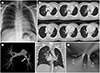
Chest radiography showed a mild leftward shift of the mediastinal structures with a reduction in the size of the left hemi-thorax and a downward-displaced left hilum (Fig. 1A).
Multidetector CT scan was performed on a 64-slice CT system (Discovery CT 750HD; GE Healthcare, Milwaukee, WI, USA) to obtain precontrast and postcontrast CT scans with iodinated non-ionic contrast. Contrast-enhanced chest CT revealed a mediastinal shift towards the left hemi-thorax, due to volume loss of the left lung (Fig. 1B). There was no left upper lobe parenchyma, bronchus, artery, or vein. Mild compensatory hyperinflation of the right lung was also observed. The main pulmonary artery trunk was enlarged in width and there was no left upper lobar artery (Fig. 1C). There was no vascular anomaly in the right hemithorax. The trachea divided into right upper lobar bronchus, bronchus intermedius, and left main bronchus at carina, which exhibited a unique trifurcation configuration (Fig. 1D). Bronchus intermedius showed focal narrowing at the site of origin. Tracheobronchial anomaly was explicitly displayed on virtual bronchoscopic image (Fig. 1E).
There was mild pulmonary hypertension (39 mm Hg) on transthoracic echocardiogram without any other congenital heart disease. He was diagnosed with a case of agenesis of the left upper lobe with tracheal trifurcation. After receiving conservative treatment at our hospital, the patient was discharged without further evaluation because of improved patient symptoms.
DISCUSSION
Pulmonary underdevelopment can range from mild pulmonary parenchymal hypoplasia to total bronchial tree and parenchymal agenesis. This developmental anomaly was first described by de Pozzis in 1673; the first proposed classification of pulmonary underdevelopment was introduced by Schneider in 1912 (Class I, Agenesis, total absence of bronchus and lung; Class II, Aplasia, rudimentary bronchus and lung; Class III, Hypoplasia, bronchial hypoplasia and variable but reduced amount of lung tissue) (2). Agenesis of one or two lobes of a single lung is an extremely rare condition; the exact incidence of this anomaly is unknown. According to Berrocal et al. (3), the most frequently affected lobe is the left upper lobe. Solitary lobar agenesis goes undetected or is incidentally detected in majority cases. However, patients with pulmonary agenesis may exhibit several accompanying congenital abnormalities, including cardiovascular diseases, as well as spinal, genitourinary, and musculoskeletal pathologies (4). Our patient exhibited left upper lobar agenesis with associated tracheobronchial anomaly - tracheal trifurcation.
Tracheal trifurcation is the most dramatic form of right tracheal bronchus, in which right upper lobe bronchus is displaced at the carina, with separate bronchus intermedius and left main bronchus originating at the same level (5). In our patient, tracheal trifurcation was distinct, revealing three even-sized bronchi at the subcarinal level, due to a reduction in the size of the left main bronchus that resulted from the absence of the left upper lobar bronchus. Patients with tracheal trifurcation typically are asymptomatic and the trifurcation is detected incidentally on imaging (6). Some patients may present with recurrent respiratory tract infection, which may progress to bronchiectasis and chronic bronchitis. Tracheal trifurcation can be diagnosed easily and rapidly by conventional CT and by three-dimensional (3D) reconstruction. It is important to identify this anatomical variant in patients requiring intubation (7). Because, the recognition of tracheal anomaly before induction of intubation can be helpful for determining optimal positioning of the endotracheal tube and it can provide accurate diagnosis especially when he/she suffers from complications due to surgery or tracheal intubation, such as lobar collapse.
Lobar agenesis, lobar collapse, and Swyer-James syndrome can be considered when the unilateral lung volume is reduced or mediastinal shifting on chest X-ray.
CT is a helpful diagnostic modality for identifying lobar agenesis. In lobar agenesis, the volume of the affected hemi-thorax is reduced; mediastinal shift towards the affected site and herniation of the contralateral lung can be observed (8). 3D reconstruction provides more and easier visualization of tracheobronchial anomaly than conventional CT and it can be particularly helpful in delineating abnormalities of the bronchi, as well as associated arterial and venous structures (49). A virtual endoscopic image can be obtained through image reconstruction; this plays an important role in characterizing bronchial anatomy without invasive bronchoscopy.
In conclusion, lobar agenesis associated with tracheal trifurcation is an extremely rare anomaly. Chest CT and 3D reconstruction may be useful in diagnosing the extent of this anomaly and evaluating associated abnormalities.
 XML Download