

 PDF
PDF ePub
ePub Citation
 Print
Print


INTRODUCTION
The fistula between gastro-pleural and gastro-pericardial space is a rare and potentially life-threatening condition. The mortality rate of gastro-pericardial fistula is known as high as 85%. The cause of gastro-pleural and gastro-pericardial fistula is variable, such as perforation of gastric ulcer, malignancy or postoperative complications (123).
There are multimodal approaches to treat advanced cancer, but the advantages are counterbalanced due to treatment-related toxicities. Recently advanced radiation technology allows for accurately targeted radiation beams, which reduces the adverse effects on the normal tissue. However, higher-dose radiotherapy can still lead to complications (4). Some studies have reported that combination of chemotherapy and radiation therapy increases the risk for fistula formation (3456). We present a case of gastro-pleuro-pericardial fistula in a patient with tongue cancer who was treated with chemoradiotherapy.
CASE REPORT
On September 2017, a 41-year-old woman presented with persistent dyspnea, cough, and sputum for the past 4 days.
One and a half year prior to the above presentation, she was diagnosed with well-differentiated squamous cell carcinoma of the tongue, with the staging of T2N0M0. She was treated with partial glossectomy and anterolateral thigh free flap reconstruction. One year later, she suffered from pleural metastasis and was treated by a palliative regimen of chemotherapy with paclitaxel and vinorelbine, and palliative radiotherapy for left posteroinferior chest wall area with a total dose of 36.5 Gy, 4 days a week, in 14 fractions in the outside hospital.
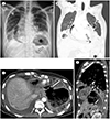
After 1 week of admission, she complained of sudden onset of chest discomfort with left lower chest pain. A chest radiograph showed a large amount of pneumopericardium, and a large cavity with air-fluid level at left lower portion, distinct from stomach air. (Fig. 1A). Chest CT demonstrated pneumopericardium and an abnormal passage between the pleura, pericardium, and stomach showing gastro-pleuro-pericardial fistula. The cavity with air-fluid level demonstrated on radiograph was identified as hydropneumothorax on CT (Fig. 1B–D).
Echocardiography demonstrated lack of the normal inspiratory collapse of the inferior vena cava, which is a sensitive echocardiographic sign of cardiac tamponade (7).
Conservative management was suggested due to her terminal status and short life expectancy. Drainage via nasogastric tube was performed. After gastric and pericardial decompression, dyspnea was improved after nasogastric tube insertion. Despite receiving treatment, the patient died a month after admission due to coagulopathy and respiratory failure.
The review of this report was exempted by the Institutional Review Board of our institution.
DISCUSSION
Pneumopericardium is a rare clinical entity of presence of air in the pericardial cavity. Cause of pneumopericardium is variable, such as chest trauma, pericardial infection, and fistula to the pericardium (8).
A fistula to pericardium can originate from any hollow organs adjacent to the pericardium. It can arise as a consequence of perforated gastric ulcer, malignancy, or postoperative complications (12389). A gastropleural fistula is a rare condition first described by Markowitz and Herter (9), as a result of intrathoracic perforation of the stomach in a hiatal hernia. A pleuropericardial fistula is also very rare. Only one case of iatrogenic pleuropericardial fistula during epicardial access of percutaneous epicardial catheter ablation for recurrent ventricular tachycardia has been reported (10).
The clinical symptom of gastropleural and gastropericardial fistula may include signs and symptoms related to pneumopericardium, pleural effusion, and perforated stomach. Epigastric and retrosternal pain radiating to the left shoulder is frequently reported with dyspnea and tachycardia (3). Pneumopericardium resulting from gastro-pleuro-pericardial fistula is even rarer. Our case report described a gastro-pleuro-pericardial fistula originated from the perforation of pleural metastasis of tongue malignancy, which was adjacent to the stomach, pleura, diaphragm, and pericardium, probably as a consequence of combined chemotherapy and radiation therapy. To the best of our knowledge, this is the second case of gastro-pleuro-pericardial fistula. The previous case report of a gastro-pleuro-pericardial fistula has been reported by Neri et al. (3). In the reported case, the diagnosis of fistula was established by chest CT and echocardiography. Our case differs from the reported one based on the cause of the fistula. In the reported case, fistula originated from a perforated gastric fundal peptic ulcer (3).
In our case, radiotherapy and chemotherapy for pleural metastasis from squamous cell carcinoma of the tongue may have predisposed our patient to develop a gastro-pleuro-pericardial fistula. Few previous cases of squamous cell carcinomas of the lungs showed fistula formation as a complication of radiation necrosis (456). Also, the study by Neri et al. (3) hypothesized that combination of radiotherapy and chemotherapy may have increased the radiation-induced endothelial toxicity, which plays a role in the formation of adhesions between the gastric fundus and the diaphragm, and led to extensive tumor vessel destruction and consequent tumor necrosis. In the setting of the previous radiotherapy, tissue damage by radiotherapy has long-term effects and various anti-angiogenic efficacies of drugs remain highly effective even in the chronic stage of long-term chemotherapy, which increases the risk for fistula formation (3456).
Recent advanced radiation technology allows for accurately targeted radiation beams, which reduces the adverse effects on the normal tissue. However, higher-dose radiotherapy can still lead to complications (4). Even though our patient was exposed to a palliative radiation dose of 36.5 Gy, which is less than the conventional dose, development of gastro-pleuropericardial fistula was noted.
In conclusion, in a patient with thoracic and respiratory symptom who has been treated with chemoradiotherapy for pleural metastasis, adequate evaluation for fistula by appropriate management is necessary. A good knowledge of typical signs of pneumopericardium and presence of abnormal passage between organs in CT could help the early diagnosis.
 XML Download