

 PDF
PDF ePub
ePub Citation
 Print
Print


INTRODUCTION
Ureteric entrapment within the sacroiliac joint (SIJ) is a very rare condition, only 5 such cases have been reported to date (12345). Almost all reported cases of ureteric entrapment within the SIJ were associated with a history of trauma (234). However, we experienced a case of unilateral ureteric entrapment within the SIJ in an elderly woman without a history of trauma. Here in we present this case along with a review of other similar cases.
CASE REPORT
A 77-year-old woman presented to our hospital with right hydronephrosis, discovered during a routine health check at outside hospital. She appeared healthy and had no history of trauma. No signs or symptoms of urinary tract infection were found on physical examination. Laboratory evaluations, including urinalysis, blood urea nitrogen, creatinine, and electrolytes were within normal limits.
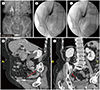
Outside ultrasonography had been performed 2 months earlier and had revealed hydronephrosis in the right kidney. Subsequently, intravenous pyelography (IVP) was performed. On IVP, the proximal ureter and renal pelvocalyx were shown to be dilated (Fig. 1A).
She was then referred to our hospital, where retrograde pyelography (RGP) and retrograde ureteric catheterization were attempted. RGP revealed kinking of the right mid-ureter at the SIJ level and dilatation of the proximal ureter (Fig. 1B). We next attempted retrograde ureteric catheterization during cystoscopy; a guide-wire could be inserted into the proximal ureter, and showed a kinking appearance, but a double-J catheter could not be inserted into the proximal ureter because the right mid ureter was entrapped at the SIJ (Fig. 1C).
Dynamic computed tomography (CT) was performed. On dynamic CT, the right kidney was shown to have hydronephrosis accompanied by right proximal hydroureter and there was no evidence of a ureter stone or solid mass along the urinary tract. The size and shape of the right kidney were normal and the enhancement pattern was also shown to be normal. In coronal and oblique sagittal images, reconstructed with the 3D software TeraRecon (Aquarius, Foster City, CA, USA; version 4.4.12), the right mid-ureter was shown to be entrapped in the right SIJ (Fig. 1D), and abrupt luminal narrowing of the ureter was seen at the portion immediately distal to the SIJ (Fig. 1E). Based on these imaging findings, our tentative diagnosis was ureter entrapment at the SIJ, and clinically differential diagnosis was ureter tuberculosis.
The patient underwent surgery in the urology department; it was confirmed that the upper ureter was severely dilated, that the mid-ureter was kinked, and that the ureter was entrapped in the SIJ of the pelvic bone. Thus, ureter excision was performed at the entrapment site, and end to end anastomosis was performed. Microscopic examination revealed chronic inflammation with fibrosis at the entrapment point. The patient recovered well, and was discharged 1 week after surgery.
DISCUSSION
Ureteric entrapment within the SIJ is very rare, with only 5 cases reported to date; these include one case reported by Otsuru et al. (1) from a cadaveric study, and there have been just 4 clinical cases reported. In 3 of the 4 clinical cases, there was a history of previous trauma; the exception is the case reported by Yeung et al. (5) In the first case, the ureter was entrapped during reduction of a pelvic fracture in a 6-year old boy (2). In the second case, the ureter was found entrapped in the SIJ during emergency exploration for ruptured bladder associated with pelvic fracture (3). In the third case, the ureter was found entrapped in the SIJ after emergency exploration for splenic injury and retroperitoneal hematoma associated with pelvic fracture (4). These cases illustrate that significant pelvic trauma, especially pelvic bone fracture, may cause ureteric entrapment within the SIJ.
Yeung et al. (5) reported bilateral ureteral entrapment in an older woman who had no traumatic or operative history, and they considered that her multiparous history or degenerative change of the SIJ predisposed her to ureteral entrapment at the SIJ.
In our case, the patient had no traumatic or operative history or multiparous history, but degenerative changes of osteophytes were seen, thus, we agree with Yeung et al. (5) that the widening caused by degenerative change may be a predisposing factor for ureteral entrapment at the SIJ.
In the case described by Yeung et al. (5), impaired renal function was observed, with elevated serum creatinine levels. Therefore, the patient was treated conservatively with bilateral percutaneous nephrostomy, and follow-up renal function tests showed normal creatinine level. Long-term management was discussed with the patient and her relatives. The option of bilateral uretero-ureterostomy was offered but declined in view of old age and fragility (5).
In contrast to the case by Yeung et al. (5), with a clinical renal problem, our patient did not show any clinical problem. We think that the hydronephrosis caused by ureteral entrapment in our case had occurred long ago; it had become a chronic problem, but did not result in complete obstruction of urine flow. We checked for signs of chronic inflammation and fibrosis at the entrapment point, which supported our hypothesis. The imaging study revealed severe constriction of the lower ureter. Ureteric tuberculosis is characterized by a thickened ureteric wall and stricture. Stricture usually involves the distal one-third of the ureter and occurs at sites of normal anatomic narrowing, such as the uretero-pelvic junction, pelvic brim, and uretero-vesical junction (6). In our urologic department, ureter excision was performed in consideration of the possibility of tuberculous ureter. After excising the entrapped ureter, end-to-end anastomosis was performed.
In our case, imaging findings by RGP and multi-detector CT facilitated diagnosis. Therefore, advances in imaging techniques could make it possible to check for hidden cases of ureteral entrapment in the SIJ.
In conclusion, when encountering cases of hydronephrosis and hydroureter above the SIJ level, it is necessary to consider ureteral entrapment at the SIJ.
 XML Download