

 PDF
PDF ePub
ePub Citation
 Print
Print


INTRODUCTION
Clear cell hidradenoma (HA) is a rare tumor of sweat glands. While the tumor was traditionally regarded to exhibit eccrine differentiation, it is now accepted that it can exhibit both eccrine and apocrine differentiation (12). HA is also known as solid-cystic HA, nodular HA, eccrine acrospiroma and clear cell acrospiroma. In spite of benign nature of the tumor, local recurrence often occurs if sufficient resection margin is not obtained at the time of excision and malignant transformation can rarely occur. Hidradenocarcinoma (HAC), which is a malignant counterpart of benign HA, can develop de novo or arise from HA.
Typical image findings of HA were recently described, but the imaging features of HAC are not yet reported. It may be because these tumors are rare and imaging studies are often omitted due to superficial location and relatively slow growing nature of the tumor. We describe ultrasonography (US) and computed tomography (CT) features of benign HA and HAC arising from HA.
CASE REPORTS
CASE 1
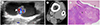
A 57-year-old female presented with a solitary palpable mass at the medial aspect of the left ankle above the medial malleolus. The mass grew slowly over 3 years. She denied pain or functional impairment. The US (Fig. 1A) showed a 2-cm, well-demarcated, mainly anechoic cystic mass with internal echogenic solid component. The inner solid component or mural nodule arising from the deep portion of the lesion showed increased vascularity on Doppler US examination. On contrast enhanced CT images (Fig. 1B), there was a mass in the subcutaneous fat layer presenting as a non-enhancing hypodense cyst with peripheral mural enhancement and internal enhancing solid nodule as reflected by mixed cystic and solid appearance on the sonography. She underwent tumor excision. During the excision, the cystic component of the tumor contained brownish fluid resembling old hemorrhage. Histological examination (Fig. 1C) showed a cystic mass mixed with solid portion. On hematoxylin-eosin stain, the specimen showed polygonal and short spindle cells with multifocal fibrous stroma, and the solid portion contained vascular channels. The tumor was pathologically confirmed as HA with moderate nuclear atypia.
CASE 2
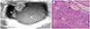
A 24-year-old female presented with a solitary palpable mass at the medial aspect of her left knee. The lesion was detected 3 years ago, and its size increased recently. The US showed a well-defined, cystic lesion with heterogeneous echogenicity in the subcutaneous fat layer (Fig. 2A). The size of the lesion was 3-cm. The lesion was composed of hypoechoic cyst with posterior acoustic enhancement and an echogenic nodule at the superficial aspect of the mass. The cystic component of the mass was not purely anechoic, suggesting intra-tumoral hemorrhage. It also contained septum like structure between the outer wall and inner echogenic nodular structure. The solid mural nodule did not reveal increased vascularity on Doppler examination. There was no evidence of distant metastasis on the positron emission tomography-CT. Excisional biopsy was performed and specimen revealed a mass with multifocal solid and cystic portions. The tumor cells showed infiltrative growth pattern invading the fibrous stroma. Multiple vascular channels were seen in the solid component of the mass with tumor infiltration within the vascular channels (Fig. 2B). The histopathology was consistent with HAC arising from HA. Additional wide excision was performed after the pathologic diagnosis, but there was no evidence of residual tumor. The patient was followed up for 2 years after the tumor excision with MR, CT, and PET-CT imaging, and there was no evidence of regional tumor recurrence or distant metastasis.
DISCUSSION
HAs are rare tumor classified as a cutaneous tumor of skin appendage. HAs are also known as solid-cystic HA, nodular HA, eccrine acrospiroma and clear cell acrospiroma. They often present as a slowly growing solitary mass, which commonly ranges from 0.5 to 3 cm in its diameter. The tumors are twice more likely to affect female than male. They are most often found in the trunk, lower extremity and head (13). Although this is a benign tumor, local recurrence often occurs when the resection margin is insufficiently obtained at the time of excision (4).
HAC is a malignant counterpart of HA. HAC can arise de novo or secondarily from preexisting HA. The rate of malignant transformation is currently not known. Malignant tumors of sweat gland are extremely rare with overall incidence of less than 0.005% (5). HAC can recur locally or metastasize distantly. Previous study reported the rate of local recurrence in the range of 14% to 20% and the rate of lymphangitic metastasis in the range of 20% to 24%. The prognosis of HAC is very poor and the 5 year post-surgical survival rate has been reported to be less than 30% (678).
The US of HA typically shows a mixed cystic and solid component similar to the present case (139). The solid component or echogenic nodular lesion commonly shows hypervascularity on Doppler exam. Increased vascularity in the solid mural nodules is due to the presence of multiple vascular channels confirmed on the histologic exam. Echogenicity of cystic component could be variable because of hemorrhage (13). Septa in the cystic component may reflect the chronicity of the tumor. Recurrent hemorrhage and/or inflammation may account for thickening of outer wall and intra-tumoral septa.
CT image findings are not specific, but it correlates well with the US features. A well-demarcated, cystic and solid lesion is typical as the current case, and they may show enhancing wall and internal solid mural nodule (1).
To our knowledge, the imaging features of HAC have not been previously reported. In our cases, differentiation of HAC from benign HA by imaging examination was difficult. It has been known that differentiation of HAC from HA is difficult both clinically and microscopically (126). Therefore, we suggest performing US-guided core needle biopsy or wide excision of tumors for any HA considering malignant potential. Further imaging evaluation for distant metastasis can be recommended to any HAC after histologic diagnosis.
The differential diagnosis of HAs includes hemangioma, lymphangioma, lymphocele, ganglion cyst, sebaceous cyst, epidermal inclusion cyst, and trichillemmal tumor. However, multiseptated appearance, which is different from the sonographic findings of HA, can easily suggest the diagnosis of hemangioma or lymphangioma (19). Even though lymphocele can be another differential diagnosis of cystic lesion, clinical history such as history of previous surgery can support the diagnosis. A ganglion cyst can also be a differential diagnosis of cystic lesion with septation. However, ganglion cysts occur nearby the joint space and thin stalks communicating with the joint space can be identified. Although epidermal inclusion cyst can present as a well-defined, mild echogenic mass like HA, internal echogenic debris can support the diagnosis of epidermal inclusion cyst. Trichilemmal tumor can present as a cystic and solid mass similar to HA. Intratumoral calcification, which is not common in HA, can be seen in trichillemmal tumor (10).
Given the rarity of these sweat gland origin tumors and lack of well-established imaging findings of HAC, we present two cases of benign HA and HAC with US and CT findings.
 XML Download