

 PDF
PDF ePub
ePub Citation
 Print
Print


INTRODUCTION
Meningiomas account for 24–30% of primary intracranial tumors diagnosed in the USA (1). The majority of meningiomas are benign, slow growing tumors with good prognoses. However, papillary meningioma (PM) is an aggressive histological variant of meningioma, accounting for 1.0–2.5% of all intracranial meningiomas diagnosed (1). According to the 2016 revision of the World Health Organization (WHO) tumor classification system (2), PM is pathologically identified as Grade III in cases where a perivascular or pseudopapillary pattern is present (3). At the time of writing, only a limited number of PM cases have been reported. We report a case of PM in a 50-year-old woman and discuss its imaging findings.
CASE REPORT
HISTORY AND EXAMINATION
A 50-year-old woman presented with a history of headache, nausea and vomiting lasting for 2 weeks. Neurological examination did not reveal any focal neurological deficit, and her past medical history was otherwise unremarkable.
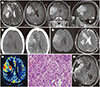
The contrasted brain MRI showed a macrolobulated margin, focal prominent inward infiltrating portion, and moderate surrounding brain edema. The mass was located at the right perisylvian inferior frontal convexity, but localization of lesion origin as intra-axial or extraaxial was difficult. The mass, which was predominantly solid with several intratumoral microcysts, measured 5.1 × 4.7 × 5.2 cm and showed mass effect compressing the right lateral ventricle. Midline shifting to the left side was also seen. Additionally, the mass showed both encasement of the right frontal middle cerebral artery branches, and peripheral engorged veins. The lesion showed heterogeneous intermediate signal intensity on T1 and T2 weighted images, and post-contrast heterogeneous enhancement (Fig. 1A). A CT scan revealed a macrolobulated, and heterogeneous enhancing mass at the right perisylvian inferior frontal convexity, with surrounding brain edema (Fig. 1B). The lesion demonstrated restricted diffusion (Fig. 1C), and high relative cerebral blood volume (rCBV) with an inward high rCBV portion (Fig. 1D).
OPERATION
A right craniotomy and tumor debulking procedure was performed. Intraoperatively, the tumor was very firm and hard, and the tumor margin showed severe adhesion with brain tissue and invasion of brain parenchyma.
PATHOLOGIC FINDING
The histopathological examination of the resected tissues revealed an increased cellularity, small cells with high nuclear-cytoplasmic ratio, and spontaneous or geographic necrosis. A perivascular pseudopapillary pattern was also seen in the resected tissue (Fig. 1E). The cells were mitotically active (mitosis 7–9/10 HPF), and prominent nucleoli were not observed. Immunohistochemistry revealed tumor cells positive for epithelial membrane antigen and vimentin.
POSTOPERATIVE COURSE
The patient was later referred to an oncology unit for radiotherapy. Twelve months later the tumor recurred, and 21 months later the tumor worsened and leptomeningeal metastasis developed (Fig. 1F).
DISCUSSION
PM is a malignant variant of meningioma first described in 1938 by Cushing and Eisenhardt (4). They reported a papillary pattern in a meningioma showing intracerebral recurrence and pulmonary metastasis. PM is frequently seen in the supratentorial compartment, though rare locations such as the posterior fossa, jugular foramen, and oculomotor nerve have been described (5).
Patients with PMs usually manifest symptoms caused by intracranial hypertension, such as severe headache, vomiting, and blurred vision, which are notably alleviated after resection of the tumor (5). PMs are more commonly seen in males and tend to occur in younger patients (6). In contrast with typical benign meningioma (WHO Grade I), our patient's MR scan showed a macrolobulated margin, which was attributed to a highly heterogeneous distribution of proliferating cells in the PM tumor, and resulted in an imbalanced cell density, and disproportionate intratumoral pressure. In our study, the tumor showed invasion of brain parenchyma, which indicates the absence of physiological barriers between the tumor and adjacent brain parenchyma. The invaded portion showed increased cerebral blood volume value and demonstrated diffusion restriction. Intratumoral microcystic change and heterogenous enhancement were also seen in this case, potentially due to microcystic degeneration, ischemic necrosis, or hemorrhage within the tumor. In previous reports, similar to our findings, high grade meningiomas usually have unclear tumor margins, heterogeneous gadolinium enhancement, and a larger scale of peritumoral brain edema (7). According to Wang et al. (5), PM shows irregular tumor margins, heterogenous enhancement, and severe peritumoral brain edema in the absence of a peritumoral rim. The presence of a tumor cyst is an exceptional finding in meningiomas, but it has been frequently reported in PM (8) in addition to our case. Yu et al. (9), who previously reported the largest and most recent series of PMs, mentioned that the MRI features of PM are unclear tumor-brain interface, and internal heterogeneity, including cyst formation, irregular enhancement, signal voids of vessels, and marked peritumoral edema. Our study showed comparable MRI features.
Due to the rarity of PM, no clear consensus exists regarding the appropriate management of the disease. However, the combination of aggressive surgical resection and postoperative radiotherapy has emerged as a standard treatment strategy for PM (5). PM is generally associated with a high mortality rate. In fact, Wang et al. (5) reported that the 3-year mortality rate for PM was 50–63.6%. In addition, local recurrence of PM has been reported in 56.5% of cases, which is significantly higher than the 10-20% recurrence rate reported for conventional meningiomas (5). The lung is the most common site of extracranial metastasis (5/10, 50%) (10).
PMs need to be differentiated from other intracranial tumors because they are malignant and have the potential for extracranial metastasis. Their timely detection could prevent local and distant metastasis, and the mortality or morbidity associated with it. The imaging findings mentioned above may serve to aide diagnosis for PM.
 XML Download