

 PDF
PDF ePub
ePub Citation
 Print
Print


INTRODUCTION
Breast cancers can be detected during screening before clinical symptoms appear by mammography and ultrasonography (US) (12), whereas symptomatic breast cancers tend to be larger and have increased likelihood for metastasis (3). The early detection of breast cancer is vitally important because it leads to the performance of breast-conserving surgery, improved treatment outcomes, and decreased mortality (45). To overcome the limitations of cancer screening by mammography, there are more studies utilizing US and magnetic resonance imaging. Due to this trend, the rates of asymptomatic early breast cancer detection have increased.
US has progressively increased in its importance as a modality for diagnosing breast cancer. Previous studies have shown that breast US has high sensitivity (98.9–99.5%) with variable specificities (37–43.7%) in diagnosing breast cancer, making it a valuable adjunct to mammography (2678910). With advances in high-resolution imaging technology, currently, breast US allows for improved rates of detection of small breast lesions. Key research regarding small breast cancers has been conducted in the past: Yoo et al. (11) studied the detectability and imaging characteristics of axillary metastasis in patients with small breast cancers (≤ 10 mm) (11), and several other studies analyzed the US features of small breast cancers (≤ 10 mm) (1213). However, these studies did not mention details regarding the US features of small breast cancers, except that most were masses (< 88%) rather than parenchymal heterogenic lesions (< 10%).
In the 8th edition of the American Joint Committee on Cancer (AJCC), the T staging of breast cancer is determined based on the size of the invasive component (14). The T1a stage is defined as < 5 mm. Until now, there has been no research on the comprehensive study of the US features of large breast cancer compared with small breast cancer < 5 mm, including the clinical and pathological characteristics. We hypothesized that the US features of small breast cancers (pathologic size ≤ 5 mm) are different from those of larger breast cancers and are less suspicious for malignancy.
Therefore, the purpose of this study was to identify differences in US feature, clinical and pathological characteristics including immunohistochemical (IHC) characteristics between small breast cancer (pathologic size ≤ 5 mm) and large breast cancer (> 5 mm).
MATERIALS AND METHODS
PATIENTS AND LESIONS
This retrospective study was approved (2016-11-045) by the Institutional Review Board of Mokdong hospital of Ewha Womans University, who waived informed consent. From January 2013 to March 2015, 591 patients with 635 invasive breast cancer lesions underwent breast surgery in our clinic. They were evaluated by preoperative US, including color Doppler imaging studies. Among them, 32 cases were excluded due to the prior use of neoadjuvant chemotherapy, and 75 pathologically confirmed ductal carcinoma in situ (DCIS) lesions were also excluded. In total, 528 invasive breast cancer lesions in 475 patients were included in this study. US-guided core needle biopsies (CNBs) or localizations were performed on all lesions prior to the surgeries. The final standard references were based on surgical pathologies.
US DATA ACQUISITION AND ANALYSIS
Bilateral whole breast US examinations, including Doppler scans, were performed by one of four board-certified radiologists (E.S.C., J.E.L., J.H.K., and J.C.) with 7–27 years of experience in breast US imaging. US was performed using 7.5–15 MHz linear-array transducers (Aixplorer system, Supersonic Imagine, Aix en Provence, France; iu22, Philips Healthcare, Bothell, WA, USA; GE LOG-IQ 9, GE Medical Systems, Milwaukee, WI, USA). The four radiologists independently analyzed and recorded US features according to the US American College of Radiology Breast Imaging-Reporting and Data System (ACR BI-RADS) (15), which was retrospectively analyzed for this study.
The US findings were analyzed according to the US BI-RADS lexicon, except for calcification (Table 1) (15). Although calcifications are included in the US BI-RADS lexicon, they are poorly characterized with US; thus, we only focused on small invasive breast cancers (15). Vascularity features were defined by three degrees (absent, minimal, and marked) (Table 1) (15).
US-GUIDED CNB AND LOCALIZATION
The US-guided localizations were executed on non-palpable lesions. The number of core samples per lesion obtained ranged from four to six. The preoperative US-guided localizations were performed using a 5-cm 20-gage Kopans spring hook wire (Cook Medical, Bloomington, IN, USA). Localizations were used to determine the scope of the surgery prior to surgery. The pathology reports of the localized lesions were produced separately.
IMMUNOCHEMISTRY AND HISTOPATHOLOGY REVIEWS
All lesions were resected surgically and reviewed by pathologists. Pathologic reports were retrospectively reviewed, and the following data were recorded for analysis: pathologic size, histologic grade, nuclear grade, and presence of either an extensive DCIS component or a lymphovascular invasion. Immunohistochemical results, including estrogen receptor (ER), progesterone receptor (PR), human epidermal growth factor receptor 2 (HER-2), and Ki-67, were reviewed and recorded. The scores of ER and PR status were classified as positive if more than 10% of the tumor cells were immunoreactive upon evaluation of 10 random microscopic fields comprising at least 1000 cells (16). The HER2 status was graded as 0, 1+, 2+, or 3+, whereas the scores of 0 and 1+ were classified as negative, and 3+ was classified as positive. In the tumors with a 2+ score, fluorescent in situ hybridization-analysis gene amplification was used to confirm HER2 status. The Ki-67 values were measured as percentages and divided into two groups (≥ 14% or < 14%) according to Ki-67 percentages (417).
STATISTICAL ANALYSIS
All statistical analyses were performed using SPSS 21.0 software (IBM Corp., Armonk, NY, USA). An independent two-group t-test was used to compare continuous variables between small breast cancers (≤ 5 mm) and large breast cancers (> 5 mm). of the categorical variables. We hypothesized that US features of small breast cancers are different than that of larger breast cancers and are less suspicious for malignancy. We compared the small breast cancers on the basis of the following US features: irregular shape, not parallel orientation, not a circumscribed margin, hypoechoic pattern, presence of posterior feature, marked vascularity, and BI-RADS category 5 to confirm that small breast cancer shows less suspicious features than large breast cancer. The Chi-squared test was used to compare categorical variables. Uni- and multivariable analyses were performed to evaluate the factors showing significance to ≤ 5 mm small breast cancers or > 5 mm large breast cancers. The Chi-squared test was used to compare multifocal/multicentric lesions and index tumors of small breast cancers. A p value of < 0.05 was considered statistically significant. Additionally, post-hoc tests were performed according to category, if they contained the above three factors, and Bonferroni correction was applied, meaning that p values < 0.0167 were considered statistically significant.
The lesions were categorized into two groups by pathologic size (≤ 5 mm or > 5 mm). According to the 8th edition of the AJCC, the T staging of breast cancer is determined based on the size of the invasive component (14). The definition of T1 stage is a tumor size below 20 mm. The T1 stage is subdivided into T1a (tumor > 1 mm but ≤ 5 mm), T1b (tumor > 5 mm but ≤ 10 mm), and T1c (tumor > 10 mm but ≤ 20 mm) (14).
After surgical resection, the immunohistochemical characteristics of multifocal/multicentric lesions were not examined independently from its index tumor, and only the immunohistochemical characteristics of the index tumor were reported. Index cancer was the original primary cancer, considered the largest invasive cancer in the breast, as defined in the 8th edition of the AJCC (14). Multifocal/multicentric lesions were considered as other multiple invasive cancers, except for the largest invasive cancer in the same or other quadrants (1819).
RESULTS
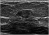
In total, 11.7% (62/528) of our cases were small breast cancers (pathologic size ≤ 5 mm). Among these, half (31/62) were index cancers, whereas the remaining half (31/62) were multifocal/multicentric cancers (Fig. 1). The margin (p = 0.002) and echo pattern (p = 0.004) were significantly different between index cancers and multifocal/multicentric cancers. The dominant features of index cancer were as follows: not circumscribed margin and a hypoechoic pattern. The dominant features of multifocal/multicentric cancers were as follows: not circumscribed margin and an isoechoic pattern. However, multivariate analysis was not possible due to the small number of cases.
COMPARISON OF CLINICOPATHOLOGICAL CHARACTERISTICS OF BREAST CANCER ACCORDING TO SIZE
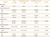
The results of the clinicopathological characteristics comparison between large and small breast cancers are shown in Table 2. Only six of the patients (9.7%) with small breast cancers showed symptoms (palpable lesion), whereas 59.9% of the patients with large breast cancers demonstrated symptoms like palpable lesion, breast pain, and nipple discharge (p < 0.001). The mean pathologic size (21.2 ± 12.27 mm/range: 6–80 mm) and mean clinical size (20.9 ± 12.97 mm/range: 5–83 mm) were similar among all the large breast cancers (p = 0.667). However, the mean pathologic size (3.4 ± 1.4 mm/range: 1–5 mm) was smaller than the mean clinical size (12.2 ± 13 mm/range: 3–50 mm) in the small breast cancers (p < 0.001).
COMPARISON OF PATHOLOGICAL CHARACTERISTICS OF BREAST CANCER ACCORDING TO SIZE
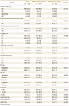
Table 3 shows that the cancer types between the large and small breast cancers were not significantly different (p = 0.714). Invasive ductal carcinoma was the most common cancer subtype for both large and small breast cancers. From the comparative result of histopathological features, only the histologic grade (p = 0.038) and nuclear grade (p = 0.040) were significantly different between the large and small breast cancers (Table 3), but this was not significant after Bonferroni correction. There was no statistically significant difference in ER, PR, HER-2, and Ki-67 between the two groups (all p > 0.05). There was no significant difference in the immunohistochemical subtypes of breast cancer between the large and small breast cancers.
COMPARISON OF THE US FEATURES OF BREAST CANCER ACCORDING TO SIZE
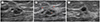
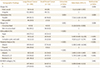
Table 4 shows the US features of the large and small breast cancers. The small breast cancers had a significantly higher rate of oval, round shape, parallel orientation; circumscribed margin; and iso/solid and cystic echo pattern, with no posterior features (all p < 0.05) as shown in Table 4 (Fig. 2). The predicting factors of small breast cancers were parallel orientation (odds ratio: 7.516), isoechoic pattern (odds ratio: 8.403), no posterior feature (odds ratio: 6.308), and minimal vascularity (odds ratio: 5.744). The small breast cancers had a higher rate of BI-RADS category 4, whereas the large breast cancers had a significantly higher rate of BI-RADS category 5.
DISCUSSION
The small breast cancers showed different US features than the large breast cancers. According to the 5th edition of ACR BI-RADS, suspicious US features for breast cancers are irregular shape, not circumscribed margin, hypoechoic pattern, and not parallel orientation (15). The results of this study, however, were different from the typical breast cancer, as the small breast cancers showed higher rate of oval, round shape, parallel orientation, circumscribed margin, iso/solid and cystic echo pattern, no posterior feature, and final assessment of category 4. Small breast cancer showed relatively low suspicion US features than large breast cancer. In our research, all of the US features were p ≤ 0.009, except those of an irregular shape (p = 0.959). Our results support the idea that small breast cancers do not show typical breast cancer features but have relatively benign US features compared with large breast cancers, mostly because small breast cancers are too small to characterize based on established classic breast cancer features. Based on our results, we conclude that the level of suspicion should be elevated in small breast lesions ≤ 5 mm, particularly if the patient has been diagnosed with breast cancer other than small US-detected lesion.
The small breast cancers had higher rates of category 4 as a final assessment, whereas the large breast cancers had higher rates of category 5. This reflects the relatively benign appearance of small breast cancers compared with large breast cancers. In a previous study, the common final assessment of breast cancers observed the mean size of target lesions to be 21 mm (range, 4–80 mm; standard deviation, 13 mm), and these lesions were categorized as BI-RADS category 5 (6). Additionally, the results of our study are consistent with the results of Korpraphong et al. (13), which showed that 84.8% of small breast cancers (≤ 10 mm) were assessed as category 4.
Regarding differences in pathologic and clinical sizes, there was an 8.8-mm gap between the pathologic and clinical size in the small breast cancers (Table 3). Seventeen of the 62 small breast cancers were markedly larger in US size (range: 17–50 mm). This discrepancy between pathologic and clinical size is probably due to the extensive DCIS component (size: 20–58 mm) seen on pathological examination in these small breast cancers. The remaining small breast cancers (45/62) were much smaller at < 10 mm in size on US. Therefore, the presence of the extensive DCIS component results in an overestimation of the size measurements during US, leading to the significant gap between pathologic and clinical size. In addition, peritumoral reactions, such as desmoplastic and lymphocytic reactions, may have contributed to the size differences, consistent with the results of a previous report by Gruber et al. (20), which showed significant overestimation in size in the US examination of breast cancers ≤ 5 mm (20).
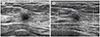
Upon further examination of the subset of six patients (9.7%) with small breast cancers (≤ 5 mm) presenting a palpable lesion, one was found to be a multifocal lesion, and the others contained DCIS components (range: 20–56 mm) (Fig. 3).
Several limitations need to be considered with this research. Owing to its retrospective nature, unavoidable selection bias may have occurred because only excised lesions with preoperative biopsy or localization were selected. Because we retrospectively analyzed the image features of breast cancer based on pathologic size, we could not consider the DCIS component and analysis of the image features of DCIS and the small invasive component of DCIS separately. Second, not all small lesions underwent localization. Pathologic reports do not contain unmarked small lesions, which may be a limitation. However, lesions were included for those which pathological correlation of localization was possible. Third, the immunohistochemical characteristics of multifocal/multicentric lesions of small breast cancers were not independently analyzed from the index cancer in these cases. Only the presence of an index mass (large breast cancer) was confirmed through immunohistochemistry. Fourth, we used several types of US devices for breast evaluation, which may have introduced bias due to different resolutions. Fifth, we did not conduct multivariable analysis to include pathological features, including clinical features and IHC subtype with imaging features. Finally, this study only included 62 small breast cancers. There is a large difference between the number of small and large breast cancers, but we did not perform further analysis using propensity score matching. Therefore, in future studies, a large population study across multiple institutions is needed to conclusively determine that the US findings of small-size breast cancers are statistically different.
In conclusion, our results show that small breast cancers have less suspicious US features than large breast cancers.
 XML Download