

 PDF
PDF ePub
ePub Citation
 Print
Print


INTRODUCTION
Acute pancreatitis is a common cause of gastrointestinal disease and associated hospital admissions (1). The incidence of acute pancreatitis varies between 4.9 and 73.4 cases per 100000 (234), and acute pancreatitis accounts for 1.9–2.5% of patients visiting emergency departments (EDs) for abdominal pain (56). In EDs, the role of advanced imaging has recently increased substantially to enhance diagnostic accuracy and facilitate treatment planning. Between 1996 and 2007, the use of computed tomography (CT) in EDs increased from 3.2% to 13.9%, which represented the highest growth rate for CT use (7). However, along with the increasing number of CT scans, unnecessary CT scans cause hospital costs and radiation exposure to patients to escalate. Therefore, it is important that CT examinations are used appropriately, to minimize unnecessary costs and risks, and improve diagnostic accuracy in EDs.
The diagnosis of acute pancreatitis requires at least 2 of 3 features: typical abdominal pain (epigastric, radiating to the back), elevated serum lipase or amylase, at levels at least 3 times greater than the upper limit of normal, or findings of acute pancreatitis on contrast-enhanced CT or magnetic resonance imaging (MRI) (89). The role of CT in patients with acute pancreatitis provides over 90% sensitivity and specificity for the diagnosis (10). CT scanning of acute pancreatitis is usually recommended after the first 48–72 h, and the utility of early CT less than 48 h remains controversial (1112131415). As complications may develop after the early period, and early CT may underestimate the degree of necrosis (11), early CT for acute pancreatitis is not routinely recommended (10). However, when patients visit EDs, physicians usually perform CT scans as early as possible, rather than waiting 48–72 h after the onset of an acute pancreatitis attack. The benefit of an early CT scan in EDs may be evaluated if its role in changing patient management strategies or facilitating urgent procedures can be established.
The most common causes of acute pancreatitis are excessive alcohol consumption and gallstones (16). Although patients with alcoholic pancreatitis are managed with conservative treatments, patients with biliary pancreatitis require biliary stone removal or cholecystectomy. Thus, the cause of acute pancreatitis affects the treatment plan for the patient. In acute pancreatitis with biliary obstruction, early diagnosis in an ED may be associated with a higher chance of endoscopic retrograde cholangiopancreatography (ERCP), resulting in stone removal and a favorable patient prognosis (i.e., prevention of recurrent attacks and potential biliary sepsis) (13). Abdominal ultrasound is useful for diagnosing gall bladder (GB) stones in patients with acute pancreatitis, but has limited value in the evaluation of obese patients, common bile duct (CBD) stones, or cholangitis (17). Contrast enhanced CT is a fast, useful modality for evaluating biliary obstructions in EDs, and may be applied to unstable patients; conversely, MRI scans are time-consuming (i.e. 30 minutes) and costly. Therefore, the purpose of this study was to investigate whether early CT scans are useful for improving the clinical management of acute pancreatitis.
MATERIALS AND METHODS
SUBJECTS
This retrospective study received ethical approval (GDIRB2016-191) from the Institutional Review Board of Gil Medical Center; requirement for obtaining written informed consent was waived. Among 140 patients diagnosed with acute pancreatitis in the ED, who underwent abdominopelvic CT from March 2015 to March 2016, 116 consecutive adults experienced their first attack of acute pancreatitis in the ED. Patient inclusion criteria were as follows: 1) initially diagnosed with acute pancreatitis (diagnostic code: K859, K852); 2) underwent abdominopelvic CT within 48 h of symptom onset; and 3) aged over 18 years. Patients who had experienced a previous attack of acute or chronic pancreatitis were excluded. Sixty patients failed to meet the inclusion criteria, and thus were excluded. The remaining 56 patients (56/116, 48.3%) with acute pancreatitis consisted of 34 men (mean age: 48.2 years; age range: 19–81 years) and 22 women (mean age: 53.6 years; age range: 18–86 years). The mean time from symptom onset to CT imaging was 12.3 h (median: 7 h; range: 2–46 h). Baseline data was collected from patients' electronic medical records; sex, age, time between symptom onset and CT examination, vital signs, serum total bilirubin, aspartate transaminase (AST), alkaline phosphatase (ALT), alkaline phosphatase (ALP), gamma glutamyl transferase (GGT), white blood cell (WBC) count, glucose, lactate dehydrogenase, and blood urea nitrogen were recorded. The gold standard for determining the etiology of acute pancreatitis was based on a combination of clinical history, laboratory findings, and imaging findings (ultrasound, MRI) and procedure. According to the disease etiologies, acute pancreatitis cases were classified into 2 groups: biliary pancreatitis and non-biliary pancreatitis.
CT TECHNIQUE
Abdominal CT scans were performed on 64-channel multi-detector CT scanners (Somatom Definition Edge, Siemens Medical Systems, Erlangen, Germany). Unenhanced-phase scans, from the liver dome to the iliac crest, were acquired first. Subsequently, contrast-enhanced scans, from the liver dome to the iliac crest or symphysis pubis, were acquired with single (portal), dual (arterial and portal), or triple (arterial, portal, and equilibrium) phases. Scans were obtained using a bolus-tracking technique (i.e., 15–25 s after the attenuation of the aorta at the thoracolumbar junction had reached 100 Hounsfield units), a fixed 72-s delay, and a fixed 180-s delay, after intravenous injections of 1.5 mL/kg of iomeprol, up to a maximum dose of 120 mL (Iomeron 300, Bracco S.p.A., Milan, Italy). The injection rate was 4 mL/s, using a power injector. The slice thickness was 3–5 mm.
CT IMAGE ANALYSIS
CT images were retrospectively reviewed by 2 abdominal radiologists (S.H.P, Y.S) with 5 and 6 years of clinical experience in performing abdominal CT with consensus who were blinded to the patients' information. Findings of pancreatitis, local complications, and biliary pancreatitis were recorded based on structured report (Table 1): 1) absence or presence of acute pancreatitis, 2) etiology of acute pancreatitis (biliary vs. nonbiliary), 3) the radiologists also described the presence of biliary sludge and/or stone, GB sludge and/or stone, GB wall thickening, transient hepatic attenuation difference (THAD), CBD wall thickening, and dilatation (> 10 mm in diameter) of CBD and scored the probability of biliary pancreatitis using a four-point scale modified from the previously described reports (1718) (Table 1). In terms of binary interpretation of etiology, where scores ≥ 2 of four-point were taken as biliary pancreatitis-positive, based on a literature review (1718) and our own preliminary CT review from October 2014 to January 2015.
URGENT PROCEDURE FOR PANCREATITIS
Urgent clinical management included ERCP within 72 h after early CT scans was assessed in acute pancreatitis patients. The presence of any of the following was evaluated: endoscopic sphincterotomy (EST), interposition of biliary stent, and stone removal from the CBD or pancreatic duct. We recorded the time interval between the CT scan and the procedure in each patient.
MORPHOLOGICAL FEATURES OF ACUTE PANCREATITIS
Morphological features of acute pancreatitis were classified as interstitial edematous pancreatitis or necrotizing pancreatitis, based on the presence or absence of necrosis, after reevaluating CT 7 days after admission (819). If the patients did not have re-evaluation CT, the feature was classified as based on initial early CT.
STATISTICAL ANALYSIS
Differences in initial Ranson's criteria, bedside index for severity in acute pancreatitis (BISAP) scores, serum total bilirubin levels, AST, ALT, ALP, and GGT values, and the presence of urgent procedures between biliary and non-biliary pancreatitis groups, were analyzed using Student's t-tests for parametric variables, and chi-square tests for non-parametric variables. The diagnostic accuracy, sensitivity, and specificity of probability of biliary pancreatitis on early CT were calculated using a four-point scale, where a score ≥ 2 was indicative of biliary pancreatitis. For all analyses, probability values (p) < 0.05 were indicative of significant differences. All statistical analyses were carried out using MedCalc Software version 14.8.1 (Med-Calc Software Inc., Mariakerke, Belgium).
RESULTS
CLINICAL CHARACTERISTICS
A total of 56 patients with acute pancreatitis were included. Patients' clinical characteristics are given in Table 2. Alcohol abuse (21/56, 37.5%) and biliary disease (24/56, 42.9%) were the 2 most common causes of acute pancreatitis. Dyslipidemia (2/56, 3.57%), drug-induced (1/56, 1.79%), and idiopathic acute pancreatitis (8/56, 14.3%) were the subsequent causes of acute pancreatitis. Patients were classified into non-biliary (n = 32) and biliary (n = 24) pancreatitis groups based on gold standard. There were no significant differences in the Ranson's criteria or BISAP scores on admission between the 2 groups (p = 0.28 and p = 0.81, respectively).
AST levels were significantly higher in the biliary than in the non-biliary pancreatitis group [239.5 ± 227.7 (20–857) vs. 76.2 ± 82.0 (14–341), p < 0.001]. The sensitivity and specificity of AST in biliary pancreatitis (AST > 120 U/L) were 58.3% and 81.2%, respectively. ALT was significantly higher in the biliary than in the non-biliary pancreatitis group [172.8 ± 132.6 (13–433) vs. 63.2 ± 54.8 (8–244), p < 0.001]. The sensitivity and specificity of ALT in biliary pancreatitis (ALT > 120 U/L) were 62.5% and 87.5%, respectively. ALP was also significant higher in the biliary than non-biliary pancreatitis group [143.8 ± 91.4 (46–362) vs. 100.5 ± 49.6 (21–278), p = 0.0269]. The sensitivity and specificity of ALP in biliary pancreatitis (ALP > 124 U/L) were 50.0% and 75.0%, respectively. There was no significant difference in the initial total bilirubin levels between the non-biliary and biliary pancreatitis groups [1.33 ± 1.83 (0.3–10.8) vs. 2.1 ± 1.83 (0.3–6.7), p = 0.126].
CT IMAGING FINDINGS
Of 56 patients, 54 (96.4%) showed acute pancreatitis on CT. Although 2 patients had typical abdominal pain and significantly elevated serum amylase, they showed normal pancreatic features on CT (Table 3).
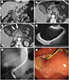
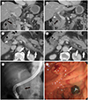
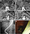
In terms of the etiology of the pancreatitis, 23 (41.1%) had biliary pancreatitis and 33 (58.9%) had non-biliary pancreatitis on early CT. The diagnostic accuracy of biliary pancreatitis on CT, compared with gold standard, was calculated using a 4-point scale. In terms of binary interpretation, where scores ≥ 2 were taken as biliary pancreatitis-positive, sensitivity, specificity, and diagnostic accuracy on CT were 91.7% (22/24), 96.9% (31/32), and 94.6% (53/56), respectively. Three patients were incorrectly assessed on CT, constituting 2 false-negatives and 1 false-positive. The 2 false-negatives (Fig. 1) were scored as 1, showing only 1 positive finding (mild CBD wall thickening); however, there was a large amount of GB sludge on endoscopic ultrasound (EUS), and they were finally confirmed as having biliary pancreatitis. The 1 false-positive was scored as 2, showing mild GB wall thickening, and a high-density lesion in the distal CBD on non-enhanced CT, suggestive of CBD sludge; however, there was no sludge on EUS. CBD wall thickening (15/24, 62.5%) was the most common finding of biliary pancreatitis, while GB wall thickening (12/24, 50.0%) was the second most common finding (Table 4). Five patients had CBD stones (Fig. 2) and 1 patient had suspected CBD sludge on CT. Eleven patients showed dilated CBD. Eight showed GB stones with GB wall thickening and THAD indicative of acute cholecystitis (Fig. 3). Five patients with GB wall thickening and THAD without GB stones on CT underwent EUS (4 patients) and abdominal US (1 patient), and were finally diagnosed with GB stones.
URGENT PROCEDURE FOR PANCREATITIS
There was a significant difference in the urgent procedures used between non-biliary and biliary pancreatitis groups (0 of 32 vs. 17 of 24, p < 0.001). Among the 24 biliary pancreatitis patients, 17 (70.8%) underwent urgent ERCP within 72 h of admission (mean time interval between CT and ERCP: 25.5 ± 19.8 h; range: 2–67 h). Of the 17 patients, six patients showed biliary stone/sludge on both CT and ERCP, four patients had radiolucent biliary stones (i.e., invisible biliary stone on CT, but visible stones during ERCP), six patients had small amounts of biliary sludge in ERCP only (i.e., invisible biliary sludge on CT), and one patient had passed CBD stone in ERCP (i.e., invisible CBD stone on either CT or ERCP). Of the 17 patients, 15 underwent EST, 9 underwent interpositions of biliary stents, 8 underwent CBD stone removal, 2 underwent infundibulostomy, and 1 underwent removal of a pancreatic ductal stone (Table 5).
MORPHOLOGICAL FEATURES OF ACUTE PANCREATITIS
Of 56 patients, 11 patients underwent re-evaluation CT after 7 days. Eight (14.3%) showed necrotizing pancreatitis (4 patients, re-evaluation CT only; 4 patients, early CT and re-evaluation CT both). Forty-six (82.1%) showed interstitial edematous pancreatitis (3 patients, both; 43 patients, early CT).
DISCUSSION
Our study showed that early CT scans can change the management of acute pancreatitis by identifying candidates eligible for urgent procedures for biliary pancreatitis. Among 24 patients with biliary pancreatitis, early CT showed a high diagnostic sensitivity (22/24, 91.7%) and 17 underwent urgent ERCP within 72 h (17/24, 70.8%) of admission, resulting in stone removal and favorable outcomes in terms of prevention of recurrent attacks. There were no urgent procedures for non-biliary pancreatitis in our study. These results suggested that non-biliary pancreatitis, including that caused by alcohol abuse, is not indicated in early CT images because of an unchanged treatment plan and underestimation of the presence and amount of necrosis. Previous studies have reported controversy about the usefulness of early CT scans for acute pancreatitis (11121314). A few studies reported that early CT scans in acute pancreatitis did not provide additional information to change the management (1012), insisting that CT for acute pancreatitis should be performed on patients in whom the diagnoses is unclear, who show no clinical improvement within the first 48–72 h after hospital admission, or to evaluate complications. However, other studies have reported the usefulness of early CT in terms of favorable patient prognoses (1415). Casas et al. (15) validated early CT as a good indicator of prognosis, and Koo et al. (14) emphasized the benefit of CT for the early identification of gallstones and biliary dilatation to induce a significant positive impact on outcome. Our findings are consistent with those of previous studies, as in patients with suspected acute biliary pancreatitis, early CT is useful for diagnosis and making appropriate procedures earlier. On the other hand, acute pancreatitis accounts for 22–73.4 cases per 100000 of the population and a ratio of gallstone to alcohol etiology of about 1–3 in western European countries and the USA, recently (20). In Korea, the annual incidence is between 15.6–19.4 cases per 100000 people and a ratio of gallstone to alcohol etiology of about 1.1 (21).
Diagnosis of acute biliary pancreatitis is crucial because patients with biliary pancreatitis require different management from those with non-biliary pancreatitis. In the literature, serum biochemical markers, such as AST, ALT, ALP, and total bilirubin, are correlated with biliary pancreatitis and CBD stones (222324). ALT is known to be useful in differentiating biliary pancreatitis from other causes of pancreatitis with high specificity but low sensitivity (18). In our study, AST and ALT were significantly higher in the biliary group than in the non-biliary pancreatitis group. Our results also showed that AST, ALT, and ALP have relatively high specificity (75.0–87.5%), but low sensitivity (50.0–62.5%). These results suggested that serum biomarkers are helpful, but insufficient, for the prediction of biliary pancreatitis. Early CT scans showed high sensitivity (91.7%) and specificity (96.9%) for the diagnosis of biliary pancreatitis, based on four-point scale scores in our study. CBD and GB wall thickening were the most common findings (62.5%, 50%, respectively) of biliary pancreatitis on CT. However, the findings of GB sludge and/or stone and CBD sludge and/or stone were relatively few (33.3%, 25.0%, respectively) in our study. Several patients had radiolucent biliary stones and small amounts of biliary sludge during ERCP (i.e., invisible biliary sludge on CT). A study of choledocholithiasis on CT has reported moderate sensitivity (60–87%) and high specificity (97–100%) (252627) and is known as a weak point of the CT scan. However, the combination of 2 or more of the findings, i.e., biliary sludge and/or stone, GB sludge and/or stone, GB wall thickening, THAD, CBD wall thickening, and dilatation of CBD complement the relatively low detection rate of choledocholithiasis on CT for the diagnosis of biliary pancreatitis.
Acute biliary pancreatitis is caused by transient obstruction of the bile and pancreatic ducts, resulting in increased within the pancreatic duct (28). For stone extraction and biliary decompression, ERCP is recommended in acute biliary pancreatitis with cholangitis or CBD stones or dilatation (29). Urgent ERCP decreases the rate of complications in severe biliary pancreatitis (30). However, ERCP is an invasive procedure, and routine ERCP in every biliary pancreatitis patient is controversial (31). Clinically, it is important to find possible candidates for urgent ERCP and findings indicative of ERCP can be seen in early CT images with patients suspected of early biliary pancreatitis.
Our study had some limitations. First, our data come from a single institution in an Asian population. Thus, our results might not be applicable to patients with different races. Nevertheless, this study would serve as a first step toward a further study. Second, our results were limited by the retrospective design and the relatively small number of patients with biliary pancreatitis. Although we recruited patients based on our inclusion and exclusion criteria, a selection bias may have resulted from the study design. As we reviewed patients' histories based on electronic medical records, there is a possibility that some findings were omitted from the records. Second, our study did not compare biliary pancreatitis with early CT to biliary pancreatitis without early CT. Third, our study did not evaluate whether early CT scans affect hospital stay, complications, and long term morbidity of acute pancreatitis. We suggested that early CT can help in identifying candidates for urgent procedures. However, a randomized controlled trial, comparing prognoses with and without early CT scans is required to demonstrate the utility of early CT in acute biliary pancreatitis.
In conclusion, early CT scans are useful for diagnosing acute biliary pancreatitis, and help to identify candidates for urgent procedures facilitating ERCP and resulting in biliary stone removal. Early CT can be performed when patients with suspected acute biliary pancreatitis visit EDs.


 XML Download