

 PDF
PDF ePub
ePub Citation
 Print
Print


INTROCUCTION
Nonalcoholic fatty liver disease (NAFLD) is a condition similar to fatty liver disease observed in alcoholic individuals and is seen in people who do not consume alcohol [1]. NAFLD has a wide range of clinical features ranging from non-inflammatory steatosis in hepatocytes to non-alcoholic steatohepatitis (NASH) [1]. Patients with NASH exhibit increased risk of developing cirrhosis or hepatocellular carcinoma over time [23]. The prevalence of NAFLD in the pediatric age group is approximately 10%, although this rate can increase to 40–70% in obese children [4]. Obesity is considered to be the most important risk factor of NAFLD [5]. Although the pathophysiology of NAFLD has not been fully elucidated, the two-stroke theory proposed by Day and James [6] is the most common hypothesis on this subject. According to this theory, the first hit involves an imbalance in fatty acid metabolism that leads to triglyceride accumulation in the hepatocytes (hepatic steatosis) and the second hit involves generation of oxidative stress and cytokines which results in liver cell damage, inflammation, and ultimately, fibrosis development [7].
Adiponectin is an adipokine that is specifically expressed in adipose tissue and directly sensitizes the body to insulin. Adiponectin regulates glucose metabolism via its anti-inflammatory and insulin-enhancing effect [8]. Adiponectin has been speculated to play an important role in the etiopathogenesis of obesity and fatty liver disease. In order to increase the metabolism of fats and glucose in obesity, the body tries to increase the adipocyte count and the adiponectin level released from adipocytes [9]. In overweight patients, muscles become resistant to insulin and adipocytes stop releasing adiponectin. Serum adiponectin levels decrease in patients with obesity, insulin resistance, and type 2 diabetes mellitus [10].
The effects of adiponectin are mediated through receptors. Adiponectin stops glucose production when it interferes with adiponectin receptor 2 in the liver and adiponectin receptor 1 in the muscle and causes glycolysis and fatty acid oxidation. C-terminal fragments of adiponectin receptors are soluble and can be detected in the plasma [11].
Adiponectin level is used in the diagnosis and prognosis of NAFLD. However, it is not a specific diagnostic marker for NAFLD. Currently, more sensitive and specific diagnostic methods are needed to diagnose NAFLD. The aim of this study was to investigate the serum adiponectin levels in obese and overweight children with NAFLD as well as obese and overweight children who have not yet developed NAFLD and study the soluble forms of adiponectin receptors based on the activity of adiponectin.
MATERIALS AND METHODS
This prospective study was performed in the Akdeniz University Medical Faculty Department of Pediatri and Department of Pediatric Gastroenterology between April 2010 to June 2011. Fifty-one children aged between 6 and 18 years, who were diagnosed with NAFLD using ultrasonography, were included in the study. Twenty normal children with similar demographic characteristics were included in the control group. The patients and their families were informed about the study, and the patients have who signed the informed consent form and declared their acceptance to participate in the study were included in the study. We excluded the patients with infections, metabolic or autoimmune diseases. Approval was received from the ethics committee of Akdeniz University Medical Faculty for this study (19.10.2010/168).
Patients were questioned for the presence of infectious diseases and drug use that could cause liver damage. Systemic examinations were performed for all patients and heights and weights were measured by the same nurse. Body mass index (BMI) (kg/m2) was calculated using height and weight measurements. Height, weight, and BMI measurements were evaluated based on age and sex using growth curves for Turkish children [12]. BMI between the 85th and 90th percentiles according to age and sex were classified as overweight and those above the 90th percentile were classified as obese [12].
Gray-scale ultrasonography (USG) examination was performed by the same radiologist using Toshiba Aplio 80 (Toshiba, Tokyo, Japan) and a multihertz convex probe that ranged from 3.3 to 5 mHz. Grading of the fat accumulation was done based on liver echography in ultrasonography images [13].
Alanine transaminase (ALT) and aspartate transaminase (AST) levels of patients included in the study were examined. Patients whose ALT level was two times higher than normal (≥80 U/L) were included in the NASH group, and patients with a value less than two times normal were included in the simple steatosis group. In addition, fasting plasma glucose, insulin, plasma cholesterol, and triglyceride levels of the whole study group were examined. Insulin resistance was calculated using the formula homeostasis model assessment (HOMA-IR) (fasting plasma insulin [μIU/mL])×fasting plasma glucose [mg/dL]/405) [14].
Blood samples were taken from the patient and control groups to study serum adiponectin and soluble adiponectin receptor 2 (soluble Adipo R2) levels simultaneously with serum ALT levels. The serum sample separated by centrifuge was maintained at −20°C at the Akdeniz University Department of Biochemistry. Adiponectin Platinum commercial kit (Bender MedSystems GmbH, Vienna, Austria) was used to evaluate serum levels of adiponectin; Enzyme-Linked ImmunoSorbent Assay was used to determine soluble Adipo R2 serum levels (Uscn Life Science Inc., Wuhan, China) using Uscn commercial kit, according to manufacturer's instructions. Serum adiponectin and soluble Adipo R2 levels were reported as ng/mL in the study.
Statistical analyses
Statistical analyses were performed using the SPSS version 15.0 (SPSS Inc., Chicago, IL, USA). Descriptive statistics were presented as frequency, percentage, mean, standard deviation, median, minimum, and maximum values. Fisher's Exact Test or Pearson chi-square test was used for the analysis of the relationships between categorical variables. In the normality test, Shapiro Wilks test was used for sample sizes smaller than 50 while the Kolmogorov-Smirnov test was used where sample size was larger than 50. Mann-Whitney U-test was used when normal distribution was predicted, while Student t-test was used for skewed distribution. In non-parametric comparison of the three disease groups, Kruskal Wallis test was used, while Mann-Whitney U-test was used as a post-hoc test in significant cases. Bonferonni correction was made for p-values. ANOVA test was used for the comparison of three groups in case of prediction of normal distribution and Tukey test was used for paired comparisons. p-values<0.05 were accepted as statistically significant.
RESULTS
A total of 71 patients, 35 males (49.3%), with a mean age of 12.91±3.43 years, were included in the study. The study group consisted of 51 patients in the NAFLD group and 20 patients in the control group. The mean age and sex was comparable between NAFLD group and control group (12.92±2.78 years vs. 12.50±3.01 years, p=0.571; 52.9% vs. 45.0%, p=0.342, respectively). In the NAFLD group, the frequency of obese patients was 72.5% (n=37) while the frequency of overweight patients was 27.5% (n=14). Half of the patients in the control group (n=10) were obese and the remaining half (n=10) were overweight. The mean BMI was 28.32±5.42 for the NAFLD group and 27.45±5.48 for the control group (p=0.550). This showed that the patient and control groups had similar demographic features (Table 1).
Table 1
The distribution of age and anthropometric measurements of the patients enrolled in the study

In the NAFLD group, mean plasma fasting glucose level was 83.17±8.51 mg/dL, mean insulin level was 16.98±8.88 U/L, mean HOMA-IR was 3.52±0.63, mean total cholesterol level was 151.10±37.40, mean low-density lipoprotein (LDL) level was 87.62±31.72 mg/dL, and the mean triglyceride level was 97.64±37.40 mg/dL. Insulin resistance was 60% in the NAFLD group. HOMA-IR, total cholesterol, LDL, triglyceride levels, and insulin resistance of obese children with NAFLD were higher than those of overweight children with NAFLD (Table 2).
Table 2
Comparison of biochemical properties of overweight and obese patients in NAFLD group
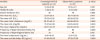
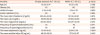
The mean ALT level in NAFLD group was 57.86±33.33 U/L and the mean AST level was 44.51±24.52 U/L. In NAFLD group, there was no difference in ALT and AST levels between obese and overweight children (69.71±39.42 vs. 53.37±30.11, p=0.167; 46.92±22.36 vs. 37.05±15.05, p=0.141). Eleven patients (21.6%) in the NAFLD group were diagnosed with NASH because of their high ALT levels. The mean age, HOMA-IR rate, and the mean total cholesterol, LDL, and triglyceride levels were similar between NASH group and simple steatosis group (who did not develop NASH) (Table 3). However, the frequency of obesity was higher in the NASH group (80% and 45%, p=0.021).
Table 3
Comparison of biochemical properties of patients with NASH and simple steatosis
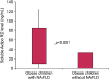
While the mean serum adiponectin level was similar between the NAFLD group and the control group, it was found below reference range in the whole study group (13.45±3.86 ng/mL vs. 10.92±4.56 ng/mL, p=0.421). The mean soluble Adipo R2 level were higher in patients with NAFLD (46.52±4.23 ng/mL vs. 8.92±3.56 ng/mL, p<0.001). In the NAFLD group, the mean serum adiponectin and the mean soluble Adipo R2 levels were comparable between the obese and overweight children (13.15±5.33 ng/mL vs. 13.61±8.56 ng/mL, p=0.321; 47.58±37.58 ng/mL vs. 41.96±32.09 ng/mL, p=0.680; respectively) (Table 2). The mean soluble Adipo R2 level was higher in obese patients with NAFLD than in obese patients without NAFLD (47.58±37.58 ng/mL vs. 10.57±11.32 ng/mL, p<0.001) (Fig. 1).
Fig. 1
Comparison of mean soluble adiponectin receptor 2 levels between obese children with nonalcoholic fatty liver disease (NAFLD) and children without NAFLD (47.58±37.58 ng/mL vs. 10.57±11.32 ng/mL, p<0.001).
The mean serum adiponectin level was lower in NASH group than in patients with simple steatosis; however, the difference was not statistically significant (12.64±5.54 ng/mL vs. 13.44±6.45 ng/mL, p=0.064). Similarly, the mean soluble Adipo R2 level was comparable between the two groups (48.63±38.29 ng/mL vs. 37.13±25.67 ng/mL, p=0.463) (Table 3).
According to the USG findings of patients with NAFLD, stage 1, stage 2, and stage 3 steatosis was observed in 18 patients (35.3%), 24 patients (47.1%), and 9 patients (17.6%), respectively. No significant difference was found in the levels of ALT, AST, HOMA-IR, total cholesterol, LDL, and triglyceride among patients staged according to ultrasonography findings. There was no significant relationship between the mean serum adiponectin level and the mean soluble Adipo R2 level as well as radiological staging (Table 4).
Table 4
Comparisons between the NAFLD stage according to ultrasonography findings and adiponectin and Soluble Adipo R2 levels

DISCUSSION
In this study, we found that NAFLD is associated with obesity in the pediatric age group. Most of the patients with NAFLD were obese while the remaining patients were overweight. Similarly, the frequency of obesity was higher in the NASH group compared to that in the simple steatosis group. Along with the increasing prevalence of obesity, NASH has become a growing health problem in the community [15]. NAFLD is becoming the most common chronic liver disease in children. In a previous study evaluating 861 obese children, NAFLD was found in 68% and metabolic syndrome in 25% patients. In this study, the researchers emphasized that NAFLD is not only a liver disease, but also an early precursor of the metabolic syndrome [16]. Since the discovery of association between obesity and hepatosteatosis, the methods for early diagnosis of NAFLD have begun to be developed.
Serum lipids levels and insulin resistance were higher in obese children than in overweight children, in NAFLD group. In addition to obesity, hyperlipidemia and insulin resistance have also been shown to be important risk factors of NAFLD [17]. After the two-hit hypothesis defined by Day and James [7] in 1998, with the determination of the importance of obesity, hypercholesterolemia, and insulin resistance in the pathogenesis of NAFLD, ‘multi-hit model’ has been developed [18].
In our study, we observed that adiponectin level was below normal in all patients included in the study. In addition, in most of the studies, adiponectin level was evaluated as μg/mL, but in our study, the results were obtained using a more sensitive unit, that is, ng/mL [19]. We found that adiponectin level was not different not only between NAFLD and control groups but also between obese and overweight children in each group. The decrease in adiponectin level observed in the control group because these children were obese or overweight. Adiponectin levels have been reported by many authors to be low in obese patients without NAFLD [2021].
Many studies have investigated the pathogenesis of NAFLD with adiponectin [2223]. Hypertrophic adipocytes produce less adiponectin in presence of insulin. The cells become more apoptotic and the cell division is delayed. As a result, plasma adiponectin level decreases. Insulin level increases in an attempt to provide more adiponectin to cells. This results in a greater increase in insulin level and a decrease in adiponectin levels [24]. Pirvulescu et al. [25] defined NASH scores using seven different parameters (BMI, ALT, AST, alkaline phosphatase, HOMA-IR, M65, and adiponectin). When the lower limit of adiponectin level was accepted as 13.5 mg/L, the sensitivity of the NASH score was 90% and the specificity was 94%. In a study by Jimenez-Rivera et al. [22], HOMA-IR and adiponectin levels were comparable between the patients with and without NAFLD, whereas there was a significant difference in serum triglyceride levels. However, in contrast to these studies, in a meta-analysis published in 2018, which reviewed 122 studies, the importance of adiponectin level in the diagnosis of NAFLD was limited (sensitivity 82% and specificity 75%). This study indicated that the gold standard diagnostic method foe NAFLD is liver biopsy [26]. With respect to NAFLD diagnosis and adipocyte activity anomaly, researchers have tried to develop new methods. For instance, C-terminal fragments of adiponectin receptors in diabetic patients were measured by plasma mass spectrometry and were found to be useful in the diagnosis of diabetic patients [11].
In our study, we found that high levels of plasma soluble Adipo R2 level might be used as a marker for predicting NAFLD in children. Soluble Adipo R2 level was found to be higher in obese children with NAFLD than in obese children without NAFLD. Adiponectin receptor 2 level was studied in the histological samples obtained from the liver and their levels were low in patients with fatty liver disease [2728]. In addition, AdipoR2 gene polymorphisms were also investigated in leukocytes obtained from peripheral blood, and it was observed that these polymorphisms affected NAFLD development [29]. This study supported that environmental factors as well as genetic factors might play a key role in disease pathogenesis.
Decreased adiponectin receptor expression in liver causes adiponectin resistance; this situation leads to the progression of the damage due to hepatosteatosis. The purpose of the treatment with peroxisome proliferator-activated receptor gamma agonists, such as rosiglitazone, is to increase the levels and sensitivity of adiponectin receptors in an organism [30]. In our study, the increase in plasma adiponectin receptor 2 level might have been due to decrease in the adiponectin level. In addition, adiponectin receptor expression may vary from individual to individual and from society to society.
In our study, no correlation was found between the clinical and laboratory findings of NAFLD staging according to ultrasonography findings. Although its common use, cheap and easy accessibility is an advantage, its dependence on the clinical experience is a significant disadvantage. In obese patients, steatosis may be overestimated due to beam attenuation by overlying fat rather than liver fat [31].
There are some limitations of our study. First, this study is a cross-sectional study. The diagnosis of NAFLD in the study group was based on ultrasonographic findings. No liver biopsy was performed on any patient, and thus, the severity of the hepatosteatosis could not be assessed by pathological findings. In addition, due to the limited number of patients, it was difficult to analyze risk factors in this group of diseases with varied etiopathogenesis.
Although there have been many studies to diagnose NAFLD in the adult, data on children and adolescents is limited. This is the first study that used the serum soluble Adipo R2 level for the diagnosis of NAFLD.
In conclusion, the prevalence of obesity in children is increasing in our country as well as globally. NAFLD is associated with obesity and metabolic syndrome and can lead to liver damage and cirrhosis if not treated at early stage. Therefore, early diagnosis and early treatment of children in risk group is important. There is a need for development of safe and easily applicable methods that can be used widely in the diagnosis and follow-up of the disease. In this context, serum soluble Adipo R2 level can be used as a non-invasive marker for the diagnosis of NAFLD. Further studies on different societies, including adult individuals, should be conducted.
 XML Download