

 PDF
PDF ePub
ePub Citation
 Print
Print


INTRODUCTION
Vitamin D deficiency accelerates bone resorption, while an increase in serum vitamin D concentrations decreases fracture risk.[1] Severe vitamin D deficiency for a prolonged period may induce osteomalacia, as mineralization of newly synthesized bone matrix is impaired.[2] A clinical state of vitamin D insufficiency may also lead to a reduction in bone mineral density of the lumbar spine and hip by decreasing intestinal calcium absorption and serum parathyroid hormones.[134]
The US National Osteoporosis Foundation and the Korean Society for Bone and Mineral Research have both recommended a daily intake of 800 to 1,000 IU of vitamin D to maintain adequate levels (30 ng/mL and more) of 25-hydroxy-vitamin D (25[OH]D). [5] Moreover, serum 25(OH)D levels at or above 30 ng/mL (75 nmol/L) are considered ideal to prevent fractures.[6] A previous study suggested that only 6.4% of Korean women have optimal levels of 25(OH)D (≥30 ng/mL), while 67.4% show deficiency (<20 ng/mL).[7] In patients recently diagnosed with osteoporosis, 69.2% of patients have a 25(OH)D deficiency (<20 ng/mL).[8] In general, supplementation with 1,000 IU of vitamin D for 3 to 4 months is required to increase serum vitamin D levels by 10 ng/mL.[9] For that reason, patients with 25(OH)D deficiency (<20 ng/mL) and insufficiency of sunlight exposure may not achieve optimal 25(OH)D levels (≥30 ng/mL), despite the recommended daily intake (1,000 IU) of vitamin D. A clinical study has shown that even with 1,000 IU of vitamin D, the vitamin D level did not reach the optimal level in osteoporosis patients.[10] Therefore, it is important to evaluate the extent to which vitamin D reaches an optimal level when treating patients with osteoporosis.
The aim of this study was to retrospectively evaluate the effect of education on sunlight exposure and additional vitamin D supplementation (1,000 IU/day) on the achievement of optimal vitamin D level in Korean postmenopausal women receiving recommended vitamin D.
METHODS
1. Study duration and patients
From March 2011, the authors have actively started patient education on the importance of exposure to sunlight to increase vitamin D synthesis. This study retrospectively analyzed 109 female patients with osteoporosis with who took the recommended amount of vitamin D (cholecalciferol 800–1,000 IU/day) during the first year (March 2011–February 2012) of active education. We measured T-score with dual energy X-ray absorptiometry (DXA) and diagnosed osteoporosis below T-score −2.5 or less. The serum 25(OH)D levels were assessed at baseline and at the end of 3 months of intervention. Of the 109 patients screened, 61 patients were included in the study. Forty-eight women who did not undergo the 25(OH)D test were excluded. The 61 patients included in the study were divided into 2 intervention groups: education only (Edu group) and education with additional vitamin D supplementation (Add group). In the Edu group, 40 patients were educated and asked to have ≥30 min sunlight exposure daily at least with the forearms and legs exposed. The other 21 patients (Add group) were educated on the importance of getting sunlight exposure for ≥30 min per day on the same condition as the Edu group and, in addition, were prescribed an additional 1,000 IU/day of cholecalciferol (for a total 1,800–2,000 IU/day). Both groups continued their prescription of cholecalciferol (800–1,000 IU/day) as part of their treatment for osteoporosis. All patients were residents of the central region at 36°N latitude in South Korea.
The occupations of the patients, as recorded from the inquiry form, were identified as 6 types, such as professional, service work, self-employment, housewife, farmer and unknown. Patients exercising more than twice a week (a total of >60 min/week) were classified as the exercise group and those not exercising as the non-exercise group.
2. Ethical approval and informed consent
The research protocol was approved by the Institutional Review Board (IRB) of the hospital (IRB No. 2016-08-021). All procedures performed in studies involving human participants were in accordance with the ethical standards of the institutional research committee, the 1964 Helsinki declaration, and its later amendments or comparable ethical standards.
3. Assessment of serum vitamin D3 status
Vitamin D3 concentration was assessed by measuring serum 25(OH)D levels at baseline and after 3 months of intervention. Vitamin D3 (25[OH]D) levels were assessed by radioimmunoassay (Dream Gamma-10, Belgium). The functional sensitivity of the assay was 4.01 ng/mL (coefficient of variation of 18.5%).
Previous studies have defined vitamin D deficiency and insufficiency as serum 25(OH)D levels <20 ng/mL (50 nmol/L) and 20 to 30 ng/mL (50–75 nmol/L), respectively. Serum 25(OH)D levels ≥30 ng/mL (75 nmol/L) are recommended for the management of osteoporosis and prevention of fractures.[1511] Based on the above criteria, the patients were classified into 3 groups: (1) deficiency (≤20 ng/mL); (2) insufficiency (20–30 ng/mL); and (3) sufficiency (≥30 ng/mL).
4. Vitamin D dose
Fourteen patients were taking alendronate-cholecalciferol (alendronate 70 mg+cholecalciferol 5,600 IU) each week, which was considered equivalent to taking 800 IU/day of cholecalciferol. The other patients (n=47) consumed cholecalciferol (1,000 IU/day) in combination with elementary calcium (500 mg/day). Of the 45 patients with baseline serum 25(OH)D <30 ng/mL, 21 received cholecalciferol (1,000 IU/day) in addition to the daily recommended dose (1,000 IU/day). Patients were prescribed FOSAMAX PLUS D (MSD Co., Seoul, Korea) as alendronate-cholecalciferol and DICAMAX 1000 (Dalim BioTech, Seoul, Korea) as cholecalciferol 1,000 IU.
5. Statistical analysis
Serum 25(OH)D levels are represented as medians (interquartile range) after normality test (Shapiro-Wilk test). The significance of the difference in 25(OH)D levels between exercise and non-exercise groups was analyzed using the Mann-Whitney U test. Differences between serum 25(OH)D levels at baseline and after intervention were analyzed with the Wilcoxon signed-rank test. Achievement rates of optimal 25(OH)D levels after the intervention were analyzed with the Fisher's exact test. All statistical data analyses were performed using IBM SPSS Statistics version 21 (IBM Corp., Armonk, NY, USA). All P-values of less than 0.05 were considered to indicate statistical significance.
RESULTS
1. General characteristics of patients and 25(OH)D status
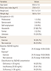
The mean age of the 61 patients was 65.6±7.6 years (range, 45–82 years). The mean height and weight were 151.7±12.4 cm and 56.9±15.4 kg, respectively. The mean body mass index was 23.6±3.3 kg/m2. The occupation was recorded as follows; housewife (60.7%), not answered (24.6%), service worker (4.9%), self-employment (4.9%), farmer (3.3%), and professional (1.6%).
The median (interquartile range) total serum 25(OH)D concentration at baseline was 25.10 (18.95–33.60) ng/mL. Serum 25(OH)D levels in exercise and non-exercise groups were 24.15 (19.53–26.65) and 25.75 (18.90–34.85) ng/mL, respectively, and showed no significant differences between the 2 groups (P=0.491). Of the 61 patients, 18 (29.5%) had vitamin D deficiency, 25 (41.0%) insufficiency, and 18 (29.5%) showed sufficiency (Table 1).
2. Increase in 25(OH)D levels after intervention
Post-intervention, the Edu group showed a significant increase in serum 25(OH)D levels in the vitamin D deficiency group (18.70 [16.52–19.00]-21.00 [20.05–29.01] ng/mL [P=0.043]), as well as the insufficiency group (25.60 [23.1–26.9]-50.51 [27.00–71.95] ng/mL [P=0.003]). In contrast, no significant increase in vitamin D levels was observed in the sufficiency group (P=0.1) (Table 2). The increase in 25(OH)D levels (mean±standard error [SE]) from baseline to post-intervention was 19.85±3.86 ng/mL in the Edu group.
Following intervention in the Add group who received education intervention as well as additional cholecalciferol (1,000 IU/day), the median (interquartile range) serum 25(OH)D level showed a significant increase from 16.60 (14.90–18.90) to 50.00 (34.20–65.37) ng/mL (P=0.003) in the deficiency group and from 25.05 (22.28–27.00) to 44.00 (41.30–58.77) ng/mL (P=0.005) in the insufficiency group (Table 2). The increase in 25(OH)D levels (mean±SE) from baseline to post-intervention was 31.73 ng/mL in the Add group.
3. Achievement rate of optimal levels after intervention
Table 3 shows the achievement rate of optimal 25(OH)D levels (30 ng/mL or above) after the intervention. In the Edu group, the proportion of participants reaching optimal levels was significantly higher in the insufficiency group than in the deficiency group (73.3% vs. 14.3%, P=0.016). In the Add group, achievement rate of optimal 25(OH)D levels was 90.9% in the deficiency group and 100% in the insufficiency group post-intervention, showing no significant differences between the 2 groups (P=0.524). The achievement rate of optimal levels was significantly higher in the Add group with non-sufficiency (25[OH]D <30 ng/mL) than in the Edu group with non-sufficiency (95.2% vs. 54.5%, P=0.003).
DISCUSSION
This study evaluated the degree of attaining the optimal vitamin D level according to the intervention in 2 groups (Edu group versus Add group), after assessing 25(OH)D levels in women under treatment for osteoporosis. Previous studies on vitamin D deficiency in Korean women are available; however, limited data are available on whether the optimal levels of 25(OH)D are maintained in patients with osteoporosis on medication. According to a previous study on 5,847 Korean female adults, only 6.4% showed adequate levels of 25(OH)D (≥30 ng/mL), and 67.4% were in a deficient state (<20 ng/mL).[7] Of all the women with an initial 25(OH)D level <30 ng/mL, 75% (55.6% in deficient group, 83.3% in insufficient group) achieved optimal levels after education for sunlight exposure and intake of cholecalciferol 1,000 IU/day for 3 months after being first diagnosed with postmenopausal osteoporosis.[8]
The serum 25(OH)D status of patients with osteoporosis indicated a deficiency in 29.5%, insufficiency in 41.0%, and sufficiency in 29.5% in this study. The rate of reaching optimal serum 25(OH)D levels was higher in patients under treatment for postmenopausal osteoporosis than those not receiving treatment, but lower than those being treated for the first time. The causes for these outcomes are unknown but appear to be associated with education of sunlight exposure and compliance with the drug dosage regimen. There were no significant differences in 25(OH)D levels between exercise and non-exercise groups. Although this finding cannot be fully explained, based on our study results, it was assumed that there were no differences with regards to the exposure time to sunlight and exposed skin area between exercise and non-exercise groups. In this study, there were very few jobs highly exposed to sunlight such as farmers.
In general, supplementation with 1,000 IU of vitamin D for 3 to 4 months is required to increase serum vitamin D levels by 10 ng/mL.[9] In the present study, serum vitamin D levels increased by 19.85±3.86 ng/mL in the Edu group and 31.73±4.82 ng/mL in the Add group. The 11.9 ng/mL difference between the 2 groups is comparable to the projected level of a 10 ng/mL increase by taking 1,000 IU/day of vitamin D. In a study on Korean diabetic patients, 25(OH)D levels were 14.6 ng/mL higher compared to those in the control group after administration of 1,000 IU cholecalciferol for 24 weeks.[12] Another study in Korean adults has reported that 25(OH)D levels increased by 17.0 ng/mL (8.5 ng/mL at a dose of 1,000 IU) compared to those in the control group after administration of 2,000 IU cholecalciferol.[13] These contradicting results seem to be attributable to the effects of varying factors (change in the amount of sunlight exposure, exposed skin area, and others) that could raise serum 25(OH)D levels. Since no assessment has been performed regarding exposure time to sunlight, exposed skin area, or drug compliance in the current study, differences between the Edu group and Add group cannot be confirmed. However, the increase in 25(OH)D levels, which is close to the estimated value of 10 ng/mL after intervention, implies that conditions may have been similar in the 2 groups.
In the present study, 29.5% of the patients with osteoporosis taking cholecalciferol (800–1,000 IU/day) before intervention had optimal levels of 25(OH)D (≥30 ng/mL). This outcome is comparable to a previous study in Spanish women where 27.7% of the patients achieved adequate 25(OH)D levels (≥30 ng/mL) after administration of 800–1,000 IU/day of vitamin D for 3 months.[14] However, the optimal 25(OH)D level was achieved in 72.5% of the patients after education, by taking a recommended dose (cholecalciferol 800–1,000 IU/day) without additional prescription of vitamin D supplement. This proportion is comparable to the achievement rate of optimal 25(OH)D levels by taking 800–1,000 IU/day of vitamin D in women diagnosed with osteoporosis for the first time.[8] These results indicate that appropriate education has a significant impact on 25(OH)D levels.
A study in Caucasian women suggests that intake levels of 800 IU/day and 1,600 IU/day are required to achieve 25(OH)D levels of 20 ng/mL and 30 ng/mL, respectively. This is based on the Recommended Dietary Allowance, which meets the nutritional requirements of 97.5% of healthy people.[15] Similarly, in the present study, the optimal 25(OH)D level was achieved in 95.2% patients in the high-dose group after taking 2,000 IU/day of cholecalciferol in combination with an educational intervention. These findings imply that a higher dose of vitamin D than the recommended 800–1,000 IU/day may be needed in order to achieve 25(OH)D levels higher than 30 ng/mL.
Maintaining an adequate level of serum 25(OH)D is important in lowering the risk of falls and fractures in postmenopausal women. In this study, the optimal 25(OH)D level (≥30 ng/mL) was achieved in 30% of women with osteoporosis without appropriate education, 72.5% following educational intervention and 95.2% after education in combination with additional vitamin D supplementation (total 1,800–2,000 IU/day). Since not all variables that could influence vitamin D levels have been considered in this study, we cannot conclude that administration of vitamin D at high doses (1,800–2,000 IU/day) is required in those with vitamin D deficiency. However, regular screening of 25(OH)D levels, despite the intake of the recommended dose of vitamin D (800–1,000 IU/day), may be helpful in maintaining optimal 25(OH)D levels in women with osteoporosis.
There are a few limitations to the study. First, as a retrospective study, this research had a limitation in classifying patients into education only and education with high-dose vitamin D. For this reason, a prospective study is warranted to confirm clinical significance. Second, since patients were recruited from a local university hospital, the study cohort is not representative of the Korean population. Third, this study was limited by the relatively small sample size. However, this investigation was meaningful in that it intended to assess the rate of reaching optimal levels of 25(OH)D and the effects of the educational intervention and additional prescription of vitamin D in patients under treatment for osteoporosis.
In conjunction with our previous findings, the optimal level of vitamin D cannot be achieved at a high rate, despite prescription of the recommended dose of vitamin D (800–1,000 IU) to patients with osteoporosis, without appropriate education. Although about 3/4 (72%) of the patients attained optimal levels after education, most of them (95%) have the potential to reach optimal levels after educational intervention and additional vitamin D supplementation (1,800–2,000 IU). Therefore, we consider that vitamin D concentration should be measured on a regular basis in order to maintain an optimal level of vitamin D, and education and additional supplementation is needed if insufficient. In addition, the results of this study provide implications for clinicians with respect to an effective means of preventing fractures for osteoporotic patients.


 XML Download