This article has been
cited by other articles in ScienceCentral.
Abstract
Leiomyomas are benign uterine smooth muscle neoplasms with varied morphology that are well known to undergo secondary changes. Cotyledonoid dissecting leiomyoma is a rare and distinct form of leiomyoma that poses a diagnostic challenge for clinicians, radiologists, and pathologists and can be confused with malignant uterine neoplasms. Only a few cases have been reported so far in the literature. Here we report a case of a cotyledonoid dissecting leiomyoma in a 60-year-old woman, emphasize its gross and histological features, and provide a review of the literature.
Keywords: Uterus, Smooth muscle, Cotyledonoid, Leiomyoma
Introduction
Leiomyomas are benign smooth muscle neoplasms that arise from the myometrium and account for almost 75% of hysterectomy cases [
1]. Such cases are mostly seen in women of reproductive age with a low incidence in postmenopausal age. Leiomyomas have numerous morphologies, among which cotyledonoid dissecting leiomyoma (CDL) is a very unusual form. Its rarity and unfamiliarity may lead to its misdiagnosis as a malignant tumor [
2]. Here we describe a case of CDL in a post-menopausal woman who presented with lower abdominal pain and third-degree uterine prolapse and present a review of the literature.
Case report
A 60-year-old woman presented with the complaint of a 1-year history of lower abdominal pain and third-degree uterovaginal descent. Her previous menstrual history was unremarkable. A vaginal examination revealed a bulky uterus with an ulceration on the anterior cervical lip. Ultrasonography revealed that the uterus was 7.5×3.4 cm in size with an endometrial thickness of 4.9 mm. Multiple uterine fibroids 2–4 cm in diameter were also noted. Both adenexa were unremarkable. A hysterectomy was performed and the specimen was sent for histopathological examination. Grossly, the cervix appeared hypertrophied and epidermidized. The endomyometrial thickness was 1.6 cm, while the endometrial thickness was 0.4 cm. Multiple fibroids up to 4×3.5 cm were noted. In addition, 2 subserosal fibroids 2 cm and 3.5 cm in diameter were seen. A cut section of the largest intramural fibroid and 1 subserosal fibroid revealed a solid, grayish-white, homogenous, and whorled appearance. The cut section of another subserosal fibroid revealed the presence of multiple grayish-white nodules (
Fig. 1A). Microscopy revealed nodules of varying sizes of uniform smooth muscles arranged in interlacing and whorling fascicles with few prominent blood vessels (
Fig. 1B–D). However, no signs of significant mitotic activity, nuclear atypia, or necrosis were seen. Based on the gross features and microscopic findings, a final diagnosis of CDL was made. Immunohistochemistry performed on CDL sections revealed diffuse positivity for vimentin, desmin, and smooth muscle actin, confirming the histopathological diagnosis of CDL (
Fig. 2). In the present case, this rare variant of leiomyoma was associated with multiple intramural and subserosal classical leiomyomas showing features of hyaline degeneration (
Fig. 3A). The endometrium was in the proliferative phase and adenomyosis was noted in the myometrium (
Fig. 3B). The cervix showed features of acute and chronic cervicitis with surface ulceration and keratinization.
Fig. 1
Gross and histopathology images of cotyledonoid dissecting leiomyoma. (A) Cut section with multiple tan white nodules. (B) Classical whorling pattern (hematoxylin and eosin [HE], ×20). (C) Nodules of varying sizes of uniform smooth muscles arranged in interlacing and whorling fascicles with few prominent blood vessels (HE, ×100). (D) Bland smooth muscles arranged in an interlacing pattern with no signs of nuclear atypia, mitosis, or necrosis (HE, ×400).

Fig. 2
Immunohistochemistry images of cotyledonoid dissecting leiomyoma showing: (A) smooth muscle actin (immunohistochemistry [IHC], ×400); (B) Vimentin (IHC, ×400); and (C) Desmin positivity in smooth muscle fibers (IHC, ×400).

Fig. 3
Histopathological images of intramural leiomyoma showing: (A) Benign smooth muscles arranged in an interlacing pattern with large areas of hyalinization (hematoxylin and eosin [HE], ×100) and myometrium; and (B) Features of adenomyosis (HE, ×40).

Discussion
Leiomyomas are the most common benign smooth muscle neoplasms of the uterus. A number of patterns of leiomyomas have been described. CDL, a very rare variant that is also commonly known as Sternberg tumor, was first reported by Roth et al. [
3] Menolascino-Bratta et al. [
4] coined the term “angionodular dissecting leiomyoma”. These tumors are frequently seen in the third to sixth decades of life. The most common complaints are lower abdominal pain and abnormal uterine bleeding. The apex case also presented with complaints of lower abdominal pain; however, no vaginal bleeding was revealed. Tumor size was typically 2–15 cm [
45]. Three types of CDL have been described in the literature. The first appears as an exophytic mass of multinodular tissue protruding from the lateral surface of the uterine cornua; resembling the placenta is called CDL. The second type is an intramural dissecting tumor that is confined to the uterus. These 2 types share similar histopathological features. The last type is pure cotyledonoid leiomyoma, which is not associated with either a parent intramural mass or intramural dissection [
6]. This case met the criteria for exophytic CDL. Microscopically, it is characterized by nodules of various sizes of uniform smooth muscles arranged in interlacing and whorling fascicles. Many blood vessels are also prominent with focal hypercellular areas. However, in contrast to malignant lesions, signs of mitotic activity, nuclear atypia, cellular pleomorphism, and necrosis are absent. Vascular invasion, capsular infiltration, and metastasis are not seen. In a few cases, perinodular hydropic changes may be prominent [
7].
A variety of other unusual patterns of uterine leiomyoma have been described, such as parasitic leiomyoma, cellular leiomyoma, symplastic or bizarre leiomyoma, epithelioid leiomyoma, intravenous leiomyomatosis, and leiomyoma with secondary changes. Some CDL appear as large fungating masses with widespread extension into the broad ligament and pelvic cavity. Due to its rarity and a clinician’s lack of familiarity, such tumors are sometimes misdiagnosed as malignancies [
8].
Gurbuz et al. [
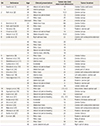
9] reported a case of cotyledonoid leiomyoma that had no intrauterine portion but had extrauterine extensions. A comparative analysis of various CDL cases reported in the literature is given in
Table 1 and Supplementary Data 1.
Table 1
Summary of the published cases of cotyledonoid dissecting leiomyoma

|
SN |
Reference |
Age |
Clinical presentation |
Tumor size (cm) maximum dimension |
Tumor location |
|
1 |
David et al. [1] |
65 |
Abnormal uterine bleed |
15 |
Uterine fundus and cervix |
|
48 |
Uterine prolapse |
12 |
Uterine fundus |
|
2 |
Roth et al. [2] |
39 |
Pelvic mass |
10.3 |
Uterine cornua |
|
41 |
Abnormal uterine bleed |
10 |
Uterine cornua |
|
23 |
Pelvic mass |
25 |
Uterine cornua |
|
Unknown |
Pelvic mass |
24 |
Uterine cornua |
|
3 |
Brand et al. [3] |
24 |
Abdominal mass |
NA |
Uterine fundus |
|
4 |
Roth and Reed [4] |
46 |
Pelvic mass |
34 |
Uterine cornua |
|
5 |
Kim et al. [5] |
26 |
Incidental |
12 |
Posterior uterine wall |
|
6 |
Cheuk et al. [6] |
55 |
Abnormal uterine bleed |
10 |
Uterine cornua |
|
7 |
Stewart et al. [7] |
58 |
Abdominopelvic mass |
16.4 |
Uterine fundus |
|
8 |
Jordan et al. [8] |
46 |
Right adnexal mass |
22 |
Uterine with extrauterine extension (all cases) |
|
46 |
Pelvic mass |
20 |
|
46 |
Pelvic mass |
10 |
|
46 |
Pelvic mass |
18 |
|
36 |
Abnormal uterine bleed |
13 |
|
34 |
Uterine mass, infertility |
18 |
|
9 |
Saeed et al. [9] |
27 |
Pelvic mass |
41 |
Uterine fundus |
|
10 |
Maimoon et al. [10] |
40 |
Urinary retention |
10 |
Uterine isthmus |
|
11 |
Shelekhova et al. [11] |
73 |
Uterine mass |
8 |
Uterine fundus |
|
12 |
Gurbuz et al. [12] |
67 |
Incidental |
10 |
Uterine cornua |
|
13 |
Weissferdt et al. [13] |
52 |
Abnormal uterine bleed |
6.2 |
Uterine fundus |
|
14 |
Raga et al. [14] |
33 |
Abdominal pain |
6 |
Lateral part of uterus |
|
15 |
Driss et al. [15] |
47 |
Pelvic mass |
25 |
Uterine with extrauterine extension |
|
16 |
Preda et al. [16] |
41 |
Uterine mass |
9 |
Left and posterior uterine wall |
|
17 |
Fukunaga et al. [17] |
56 |
Constipation |
30 |
Posterior uterine wall |
|
47 |
Abdominal pain |
26 |
Posterior uterine wall |
|
36 |
Abnormal uterine bleed |
4 |
Posterior uterine wall |
|
35 |
Abdominal pain |
18 |
Lateral uterine wall |
|
18 |
Gezginç et al. [18] |
57 |
Pelvic pain |
2.5, 4.5 |
Intrauterine, lateral uterine wall |
|
19 |
Agarwal et al. [19] |
52 |
Abnormal uterine bleeding |
10 |
Uterine cornua |
|
20 |
Ersöz et al. [20] |
51 |
Abnormal uterine bleeding |
8.5 |
Subserosal |
|
21 |
Roth et al. [21] |
33 |
Abnormal uterine bleeding |
6.5, 13.5 |
Posterior uterine wall |
|
22 |
Tanaka et al. [22] |
36 |
Incidental |
10 |
Posterior & lateral uterine wall |
|
23 |
Onu et al. [23] |
50 |
Incidental |
10 |
Uterine fundus |
|
24 |
Kim et al. [24] |
43 |
Abdominal mass |
13 |
Uterine with extrauterine extension |
|
25 |
Blake et al. [25] |
56 |
Abnormal uterine bleeding |
30 |
Uterine with extrauterine extension |
|
26 |
Shimizu et al. [26] |
40 |
Abnormal uterine bleeding |
10 |
Posterior uterine wall |
|
27 |
Rocha et al. [27] |
38 |
Abnormal uterine bleeding |
25 |
Uterine isthmus |
|
28 |
Xu et al. [28] |
55 |
Pelvic mass |
6 |
Posterior uterine wall |
|
43 |
Pelvic mass |
3 |
Body of uterus |
|
37 |
Pelvic mass |
30 |
Periuterine |
|
48 |
Lower abdominal pain |
6.7 |
Right wall of uterus |
In conclusion, CDL is a unique and rare variant of leiomyoma with a characteristic gross nodular appearance and microscopic features. Increasing awareness among clinicians and pathologists regarding this rare entity will prevent inappropriate diagnosis and treatment.


 PDF
PDF ePub
ePub Citation
 Print
Print


 XML Download