

 PDF
PDF ePub
ePub Citation
 Print
Print


Abstract
Background
Liver fibrosis evaluation is an important issue in chronic liver disease patients. We aimed to develop noninvasive liver fibrosis biomarkers based on transient elastography (TE, FibroScan®) through retrospective review of clinicopathological data.
Methods
We recruited 278 chronic hepatitis B patients who underwent Fibroscan and HBV DNA testing. A total of 115 HBeAg-positive and 159 HBeAg-negative chronic hepatitis B patients were analyzed. A total of 100 hepatitis C patients were analyzed. Successful fibroscan data, gamma-glutamyl transferase (GGT) to platelet ratio (GPR), platelet count, AST, ALT, international normalized ratio of prothrombin time, total cholesterol, triglycerides, bilirubin, mean platelet volume, AST to platelet ratio index, fibrosis index based on four factors (FIB-4), neutrophil to lymphocyte ratio (NLR), and NLR to platelet ratio were analyzed to determine the new noninvasive markers for assessing liver fibrosis.
Results
Elevated GPR (OR=9.1, P=0.011) and FIB-4 (OR=2.3, P=0.01) were associated with greater risk of liver fibrosis in chronic hepatitis B patients. FIB-4 (OR =6.04, P=0.005) was a risk factor for liver fibrosis in HBeAg-positive patients. FIB-4 (OR =2.371, P=0.015) and GPR (OR=33.78, P=0.003) were liver fibrosis risk factor in HBeAg-negative patients. In chronic hepatitis C patients, GGT (OR=1.033, P=0.002), triglyceride (OR=–0.990, P=0.038) and FIB-4 (OR=3.499, P=0.006) showed statistical significances. The AUCs were 0.816 in FIB-4 (P<0.001) and 0.849 in GPR (P<0.001).
Go to : 
References
1. Bravo AA, Sheth SG, Chopra S. Liver biopsy. N Engl J Med. 2001; 344:495–500.

2. Coco B, Oliveri F, Maina AM, Ciccorossi P, Sacco R, Colombatto P, et al. Transient elastography: a new surrogate marker of liver fbrosis in-fuenced by major changes of transaminases. J Viral Hepat. 2007; 14:360–9.
3. Colombo S, Belloli L, Buonocore M, Jamoletti C, Zaccanelli M, Badia E, et al. True normal liver stiffness measurement (LSM) and its determinants. Hepatology. 2009; 50:741A–2A.
4. Foucher J, Chanteloup E, Vergniol J, Castéra L, Le Bail B, Adhoute X, et al. Diagnosis of cirrhosis by transient elastography (FibroScan): a prospective study. Gut. 2006; 55:403–8.
5. Friedrich–Rust M, Ong MF, Martens S, Sarrazin C, Bojunga J, Zeuzem S, et al. Performance of transient elastography for the staging of liver fbrosis: a metaanalysis. Gastroenterology. 2008; 134:960–74.
6. Kim SU, Choi GH, Han WK, Kim BK, Park JY, Kim DY, et al. What are ‘true normal'liver stiffness values using FibroScan?: a prospective study in healthy living liver and kidney donors in South Korea. Liver Int. 2010; 30:268–74.
7. Pinzani M, Vizzutti F, Arena U, Marra F. Technology Insight: noninvasive assessment of liver fbrosis by biochemical scores and elastography. Nat Clin Pract Gastroenterol Hepatol. 2008; 5:95–106.
8. Arena U, Vizzutti F, Corti G, Ambu S, Stasi C, Bresci S, et al. Acute viral hepatitis increases liver stiffness values measured by transient elastography. Hepatology. 2008; 47:380–4.
9. Sagir A, Erhardt A, Schmitt M, Häussinger D. Transient elastography is unreliable for detection of cirrhosis in patients with acute liver damage. Hepatology. 2008; 47:592–5.
10. Marcellin P, Ziol M, Bedossa P, Douvin C, Poupon R, De Lédinghen V, et al. Non‐invasive assessment of liver fbrosis by stiffness measurement in patients with chronic hepatitis B. Liver Int. 2009; 29:242–7.
11. Lok AS and McMahon BJ. Chronic hepatitis B: update 2009. Hepatology. 2009; 50:661–2.
12. Kim SU, Seo YS, Cheong JY, Kim MY, Kim JK, Um SH, et al. Factors that affect the diagnostic accuracy of liver fbrosis measurement by Fibroscan in patients with chronic hepatitis B. Aliment Pharmacol Ther. 2010; 32:498–505.
13. Kim BK, Kim DY, Park JY, Ahn SH, Chon CY, Kim JK, et al. Validation of FIB-4 and comparison with other simple noninvasive indices for predicting liver fbrosis and cirrhosis in hepatitis B virus-infected patients. Liver Int. 2010; 30:546–53.
14. Liu DP, Lu W, Zhang ZQ, Wang YB, Ding RR, Zhou XL, et al. Comparative evaluation of GPR versus APRI and FIB-4 in predicting different levels of liver fbrosis of chronic hepatitis B. J Viral Hepat. 2018; 25:581–9.
15. Lemoine M, Shimakawa Y, Nayagam S, Khalil M, Suso P, Lloyd J, et al. The gamma-glutamyl transpeptidase to platelet ratio (GPR) predicts signifcant liver fbrosis and cirrhosis in patients with chronic HBV infection in West Africa. Gut. 2016; 65:1369–76.
16. Lin ZH, Xin YN, Dong QJ, Wang Q, Jiang XJ, Zhan SH, et al. Performance of the aspartate aminotransferase-to-platelet ratio index for the staging of hepatitis C-related fbrosis: an updated metaanalysis. Hepatology. 2011; 53:726–36.
17. Vallet-Pichard A, Mallet V, Nalpas B, Verkarre V, Nalpas A, Dhalluin-Venier V, et al. FIB-4: an inexpensive and accurate marker of fbrosis in HCV infection. comparison with liver biopsy and fbrotest. Hepatology. 2007; 46:32–6.
18. Li J, Verhaar AP, Pan Q, de Knegt RJ, Peppelenbosch MP. Serum levels of caspase-cleaved cytokeratin 18 (CK18-Asp396) predict severity of liver disease in chronic hepatitis B. Clin Exp Gastroenterol. 2017; 10:203–9.
19. Rosenberg WM, Voelker M, Thiel R, Becka M, Burt A, Schuppan D, et al. Serum markers detect the presence of liver fbrosis: a cohort study. Gastroenterology. 2004; 127:1704–13.
20. Kuno A, Ikehara Y, Tanaka Y, Ito K, Matsuda A, Sekiya S, et al. A serum “sweet-doughnut” protein facilitates fbrosis evaluation and therapy assessment in patients with viral hepatitis. Sci Rep. 2013; 3:1065.
21. Nishikawa H, Takata R, Enomoto H, Yoh K, Kishino K, Shimono Y, et al. Proposal of a predictive model for advanced fbrosis containing Wisteria foribunda agglutinin-positive Mac-2-binding protein in chronic hepatitis C. Hepatol Res. 2017; 47:E74–84.
22. Abe M, Miyake T, Kuno A, Imai Y, Sawai Y, Hino K, et al. Association between Wisteria foribunda agglutinin-positive Mac-2 binding protein and the fbrosis stage of non-alcoholic fatty liver disease. J Gastroenterol. 2015; 50:776–84.
23. Umemura T, Joshita S, Sekiguchi T, Usami Y, Shibata S, Kimura T, et al. Serum Wisteria foribunda agglutinin-positive Mac-2-binding protein level predicts liver fbrosis and prognosis in primary biliary cirrhosis. Am J Gastroenterol. 2015; 110:857–64.
24. Nishikawa H, Enomoto H, Iwata Y, Hasegawa K, Nakano C, Takata R, et al. Clinical signifcance of serum Wisteria floribunda agglutinin positive Mac-2-binding protein level and high-sensitivity C-reactive protein concentration in autoimmune hepatitis. Hepatol Res. 2016; 46:613–21.
25. Yamada N, Sanada Y, Tashiro M, Hirata Y, Okada N, Ihara Y, et al. Serum Mac-2 binding protein glycosylation isomer predicts grade F4 liver fbrosis in patients with biliary atresia. J Gastroenterol. 2017; 52:245–52.
Go to :
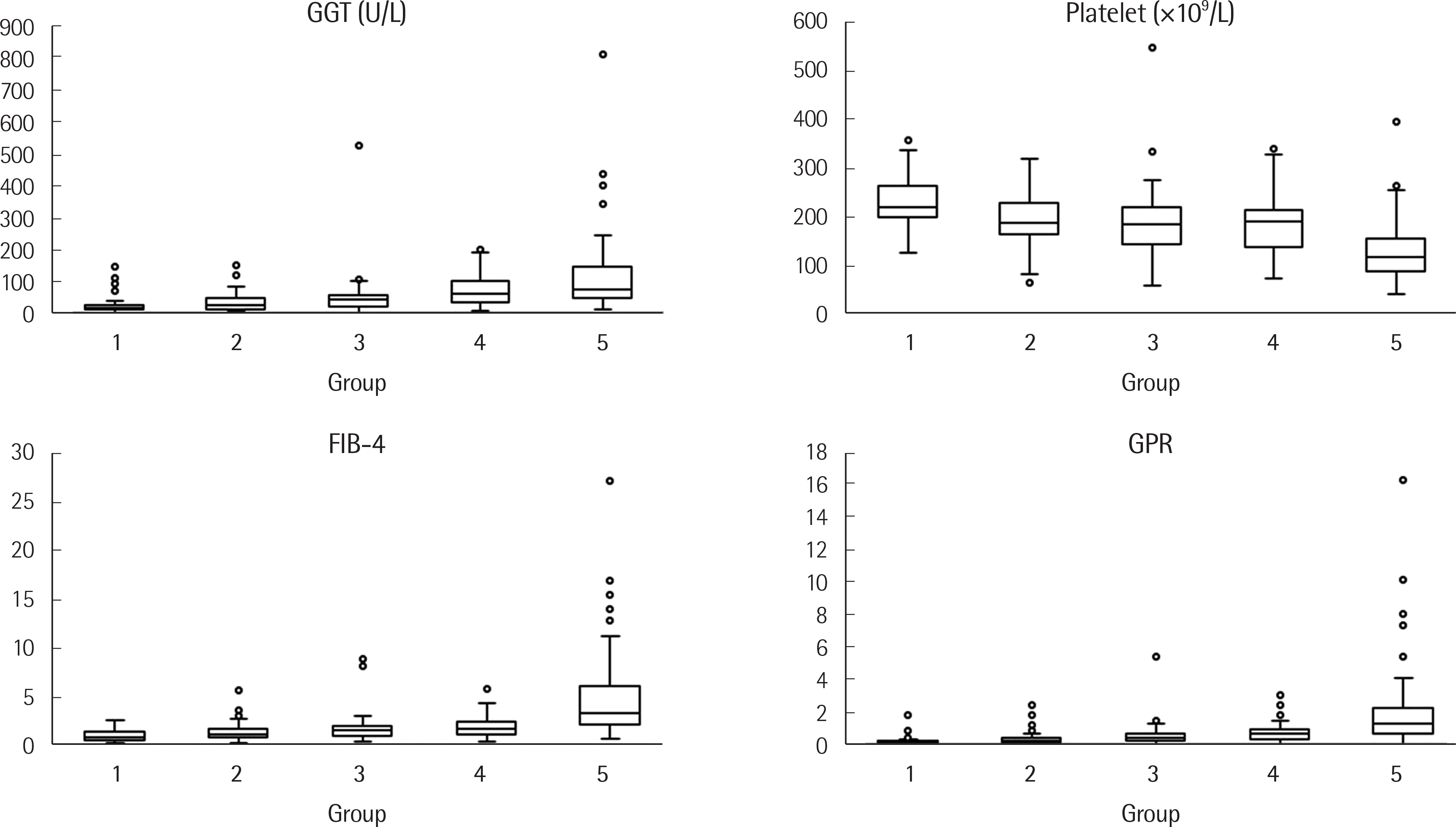 | Fig. 1.Box plot of gamma glutamyl transferase (GGT), platelet count, FIB-4 and GPR in chronic hepatitis B patients. |
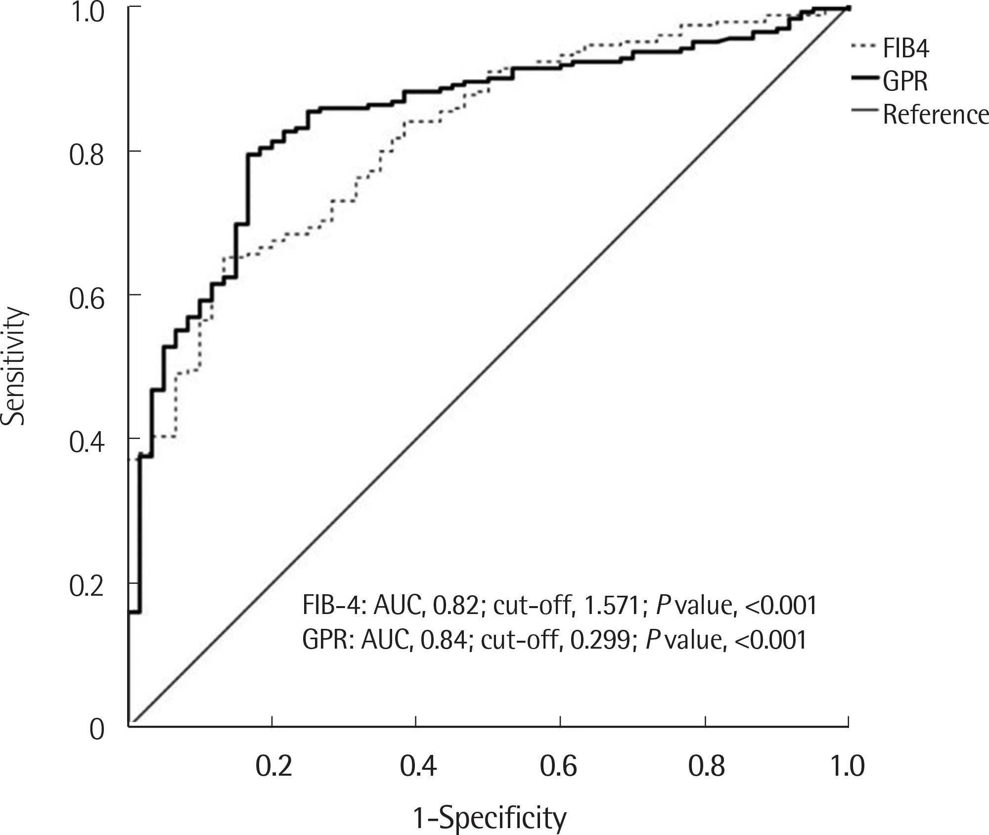 | Fig. 2.Receiver operating characteristic (ROC) curve analysis of FIB-4 and GPR for the evaluation of liver fibrosis comparing transient elastography (fibroscan) results in chronic hepatitis B patients. |
Table 1.
Summary of calculated biomarkers for liver fibrosis expectation
Table 2.
Levels of parameters according to different liver stiffness measurement levels measured by transient elastography
Abbreviations: GGT, gamma-glutamyl transferase; AST, aspartate aminotransferase; ALT, alanine aminotransferase; INR, international normalized ratio of prothrombin time; MPV, mean platelet volume; APRI, AST to platelet rati index; FIB-4, fibrosis index based on four factors; GPR, GGT to platelet ratio; NLR, neutrophil to lymphocyte ratio.
Table 3.
Diagnostic sensitivity, specificity, positive predictive value and negative predictive value of FIB-4 and GPR for liver fibrosis prediction
| Parameters | Sensitivity (%) | Specificity (%) | PPV (%) | NPV (%) | AUC | P value |
|---|---|---|---|---|---|---|
| FIB-4 (cut-off: 1.571) | 65.1 | 86.7 | 86.7 | 86.7 | 0.82 | <0.001 |
| GPR (cut-off: 0.299) | 79.4 | 83.3 | 83.3 | 83.3 | 0.84 | <0.001 |
Table 4.
Characteristics of baseline parameters according to HBeAg status
| HBeAg positive (N=115) | HBeAg negative (N=159) | P value | |
|---|---|---|---|
| Age (yr) | 45.4±11.9 | 48.3±10.9 | 0.037 |
| Sex ratio (Male:Female) | 1.7 (73:42) | 1.7 (100:59) | 0.921∗ |
| Liver stiffness measurement (kPa) | 14.751±11.156 | 10.757±10.683 | 0.003 |
| AST (U/L) | 95.8±142.3 | 63±193 | 0.124 |
| ALT (U/L) | 132.5±268 | 80.5±302.5 | 0.142 |
| GGT (U/L) | 68.3±58.2 | 63.7±94.2 | 0.644 |
| Total Bilirubin (mg/dL) | 0.9±0.55 | 1.05±1.63 | 0.344 |
| Total Cholesterol (mg/dL) | 170.5±30.8 | 172.1±35.4 | 0.696 |
| Triglyceride (mg/dL) | 94.5±41.9 | 97.4±47.5 | 0.605 |
| Platelet count (×109/L) | 174.1±73.6 | 186.4±70.1 | 0.163 |
| Mean platelet volume (fL) | 8.41±0.86 | 8.17±0.88 | 0.022 |
| Prothrombin time, INR | 1.09±0.12 | 1.07±0.16 | 0.265 |
| HBV DNA (IU/mL) | 21,028,204.8±41,665,244.9 | 591,105.9±3,276,928.3 | <0.001 |
| AST/ALT ratio | 1.11±0.71 | 1.04±0.46 | 0.282 |
| APRI | 1.747±2.596 | 1.345±5.241 | 0.449 |
| FIB-4 | 2.958±2.799 | 2.275±2.879 | 0.05 |
| GPR | 0.9±0.813 | 0.922±1.777 | 0.889 |
| NLR | 1.53±0.704 | 1.753±0.829 | 0.02 |
| NLR/platelet count | 0.011±0.008 | 0.012±0.01 | 0.309 |
Abbreviations: AST, aspartate aminotransferase; ALT, alanine aminotransferase; GGT, gamma-glutamyl transpeptidase; INR, international normalized ratio of prothrombin time; APRI, AST to platelet ratio index; FIB-4, fibrosis index based on the four factors; GPR, GGT to platelet ratio; NLR, neutrophil to lymphocyte ratio.
Table 5.
Levels of parameters according to liver stiffness measurement in chronic HCV infected patients
| LSM<5.3 kPa (N=74) | LSM≥5.3 kPa (N=26) | P value | |
|---|---|---|---|
| Genotype 1b/2a | 37/37 | 17/9 | 0.176∗ |
| Age (yr) | 53 | 58 | 0.016 |
| Male/Female | 37/37 | 11/15 | 0.499∗ |
| LSM (kPa) | 4.05 | 10.3 | <0.001 |
| AST (U/L) | 36 | 65 | <0.001 |
| ALT (U/L) | 44.5 | 82 | 0.001 |
| GGT (U/L) | 27.5 | 83.5 | <0.001 |
| Bilirubin (mg/dL) | 0.58 | 0.705 | 0.010 |
| Cholesterol (mg/dL) | 191.5 | 160.5 | 0.001 |
| Triglyceride (mg/dL) | 117.5 | 98 | 0.004 |
| Platelet (×109/L) | 237.5 | 173.5 | <0.001 |
| MPV (fL) | 7.25 | 7.9 | 0.004 |
| INR (sec) | 0.985 | 1.025 | 0.005 |
| HCV RNA | 834,000 | 1,510,000 | 0.535 |
| AST to ALT ratio | 0.86 | 0.845 | 0.786 |
| APRI | 0.405 | 1.005 | <0.001 |
| FIB-4 | 1.245 | 2.78 | <0.001 |
| GPR | 0.12 | 0.475 | <0.001 |
| NLR | 1.755 | 1.45 | 0.210 |
| NLR/platelet | 0.007 | 0.008 | 0.049 |
 XML Download